


Ferritin: Its Structure, Biological Functions, and Biomedical Applications
1. Introduction
Ferritin is a highly preserved, evolutionarily conserved protein responsible for controlling both cellular and systemic levels of available iron. Its major role is to keep stored iron in a non-toxic and easily utilized state to prevent damage to cells due to excessive amounts of free iron. This function is essential because iron is biologically indispensable, yet potentially dangerous when it accumulates outside controlled protein systems. Ferritin is not only a storage molecule but also a protective regulator of redox balance, tissue stability, and metabolic function (Yanatori et al., 2023). As a result, it has remained a major subject in multiple disciplines, including biochemistry, cell biology, pathology, and laboratory medicine (Arosio et al., 2024; Polizzi, 2026).
Clinical applications of ferritin are substantial, and serum ferritin is widely used to assess iron stores and support the diagnosis of iron deficiency. However, ferritin can be influenced by infection and inflammation; therefore, its increased levels do not automatically indicate an excess amount of iron. Studies have recently demonstrated that selecting different ferritin cutoffs for diagnostic purposes significantly affects how frequently iron deficiency is identified in primary care settings. Some studies also emphasize that ferritin should not be interpreted alone in inflammatory settings, but with other markers (Jäger et al., 2024).
2. Structural Characteristics of Ferritin
Ferritin forms a unique, spherical structure composed of 24 protein subunits, which assemble into a nano-cage capable of storing large amounts of iron in ferric form. In human ferritins, these subunits have varying combinations of heavy (H) and light (L) chains encoded by ferritin heavy chain 1 (FTH1) and ferritin light chain (FTL) (Arosio, 2024; Lee et al., et al., 2024). The H-rich form is more active in oxidation of iron and metabolic turnover, while the L-rich form is involved in the nucleation process and in long-term mineral storage (Yanatori et al., 2023). Protein data structural resources demonstrated that ferritin functions as a hollow shell with an internal iron-storage cavity, and that the H and L-chains contribute differently to iron handling. Furthermore, the L-chain entry denotes that it can host approximately 4500 ferric iron atoms within its central cavity, illustrating the remarkable efficiency of this protein system (Sudarev et al., 2023).
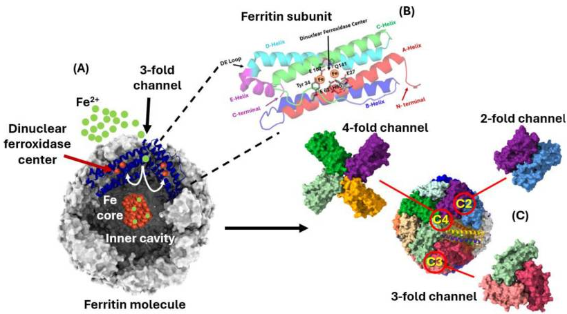
The overall structural arrangement explains the biological importance of ferritin. The encapsulation of iron in the cage creates a high-density environment where iron can be maintained in a chemically controlled manner, allowing it to be released when required for various cellular processes such as respiration, DNA synthesis, and proliferation. This highly organized structure has further gained an interest in ferritin studies in nanotechnology and drug delivery research. Ferritin is a naturally occurring protein that self-assembles into a stable nano-scale sphere, and it represents a promising tool for designing biomedical applications (Lee et al., 2022). The overall structure of ferritin showing how iron accesses the ferroxidase center via the 3-fold channel with an iron core in the middle of the ferritin cavity is shown in Figure 1 (A) below, the helix structure of one subunit is indicated in (B), and ferritin subunits interface at dimer (C2) is presented in (C), trimer (C3) and tetramer (C4) symmetry.
Figure 1: A structure of ferritin
(Arosio et al., 2024)
1. Synthesis and Regulation of Ferritin
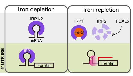
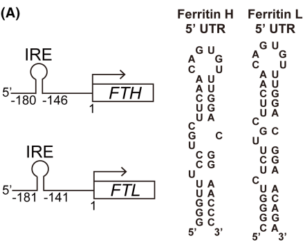
Ferritin synthesis is highly controlled because both iron deficiency and an overloaded state can be harmful (Ru et al., 2024). It is regulated by several mechanisms, including iron-dependent post-translational mechanisms. The iron regulatory protein/iron-responsive element pathway controls ferritin translation in response to intracellular iron levels, as shown in Figure 2. When iron is low, ferritin production is restricted to prevent cells from locking up their iron reserves. Conversely, when iron is high, ferritin translation increases, and the protein safely stores iron. The 5'-untranslated regions (UTRs) of FTH and FTL mRNA contain an iron-responsive element (IRE) that binds to the iron regulatory protein (IRP), regulating ferritin expression (Figure 2A). There are two forms of IRPs (IRP1 and IRP2). When the cell has low intracellular iron levels, IRP binding to IREs on ferritin mRNA inhibits mRNA translation and reduces ferritin expression. As intracellular iron stores increase, IRPs lose the ability to bind IRE. With IRPs removed from the IRE, ribosomes can translate ferritin mRNA, leading to increased ferritin expression, allowing the storage of excess iron (Figure 2B).
Recent studies have indicated that ferritin expression is influenced by oxidative stress, inflammation, and general iron-metabolic signaling, which helps explain why ferritin levels increase in many pathological states beyond classical iron overload (Polizzi, 2026; Yanatori et al., 2023). Recent research also indicates that ferritin is not simply a passive intracellular storage box. There are biologically coordinated mechanisms for its turnover, transport, and degradation; these processes influence how much iron becomes available upon stress or disease exposure. Therefore, the dynamic nature of ferritin's regulation is one of the reasons ferritin is now discussed in modern work on inflammation, infection, and programmed cell death rather than only in traditional nutrition studies or hematological contexts (Liao et al., 2025; Polizzi, 2026).
Figure 2: Regulation of ferritin synthesis
1. Biological functions of Ferritin
The primary biological function of ferritin has previously been believed to be the safe storage of excess iron. However, its biological significance extends beyond this view (Sudarev et al., 2023). By acting as a buffer for the labile iron pool, ferritin reduces the likelihood that iron can be involved in redox reactions that cause damage to cellular components such as membranes, proteins, and nucleic acids. This antioxidant protective role is especially important in active tissues and diseases associated with redox imbalance (Polizzi, 2026).. Therefore, ferritin is a major component of the cellular defense mechanism against iron-related injuries (Arosio et al., 2024). In addition, it can release iron into the cytoplasm as needed through degradation mechanisms. These mechanisms allow the ferritin-bound iron to be returned to metabolic use as required by changing cellular demands. As such, ferritin plays an important role in ferroptosis, an iron-dependent process of programmed cell death that occurs due to excessive levels of reactive oxygen species (ROS) produced during lipid peroxidation and antioxidant system failure.
Recent reviews place iron homeostasis at the center of ferroptosis biology, increasing interest in ferritin across cancer, neurodegenerative diseases, inflammatory diseases, and tissue injuries (Ru et al., 2024). Ferritin is also increasingly linked to host defense and inflammatory signaling. Evidence indicates that in infectious disease settings, ferritin may not only serve as a biomarker for inflammation but also as a participant in inflammation biology. This broad perspective explains why ferritin is often associated with disease severity and prognosis in some infections and hyper-inflammatory conditions (Liao et al., 2025).
2. Clinical Applications of Ferritin
Serum ferritin is widely used as one of the major laboratory markers for the evaluation of the body's iron status (Cancado et al., 2025; Garcia-Casal et al., 2018). Lower levels of ferritin are useful in identifying the depleted iron stores and supporting the diagnosis of iron deficiency, but the interpretation becomes more complex when inflammation is present (Cancado et al., 2025). In practice, its testing is widely used in the evaluation of anemia, fatigue, and nutritional deficiencies, along with iron-related issues during pregnancy, and excessive bleeding. More recently, studies suggest that varying ferritin cutoffs can produce highly different rates of iron deficiency diagnosis in primary care populations (Jäger et al., 2024).
One of the major limitations of ferritin is that it is also an acute-phase reactant. The World Health Organization (WHO) provided guidelines that recommend using higher serum ferritin thresholds in patients with infection/inflammation to determine if they have iron deficiency. It further recommends that elevated serum ferritin levels cannot be used as the sole indicator of iron overload. This point is significant because normal or high ferritin values may coexist with functional iron deficiency in inflammatory diseases. Therefore, the most accurate method to interpret serum ferritin results would be by correlating these levels with patient-reported symptoms, other measures of iron status, and markers of inflammation.
Serum ferritin also serves as a tool in evaluating individuals for excessive iron storage, severe infections, liver dysfunction, malignancies, and hyperferritinemias associated with inflammation. As such, the medical utility of serum ferritin extends beyond nutrition/hematology into broader disease surveillance and clinical decision making based on risk assessment (Cancado et al., 2025; Liao et al., 2025).
3. Biomedical and Technical Applications
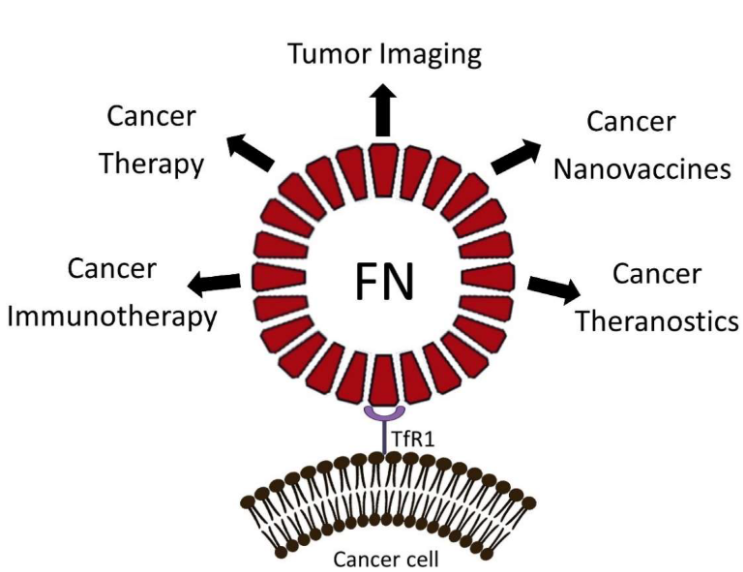
Ferritin’s ability to adapt its natural nano-cage for biomedical applications has increased significantly in recent years. Some studies describe ferritin as an endogenously derived nano-scale scaffold that possesses strong biosafety features, rapid dispersal behavior, and the potential for chemical or genetic modifications on both the cage and surface and inner cavity. Ferritin-based systems are currently explored for vaccine design, tumor targeting, imaging, and targeted therapeutic systems (Lee et al., 2022). Ferritin has a distinct advantage over most synthetic carriers as it combines structural stability with natural biological compatibility, making it a strong example of how a classical cellular protein can be transitioned to a form of advanced therapeutic design while still retaining its inherent physiological significance (Arosio et al.2022).
Figure 3: Application of ferritin in the field of Oncology
(Mainini et al., 2021)
1. Methods used to detect Ferritin
Ferritin levels are typically measured using laboratory immunoassays rather than by direct structural analysis. The most common approaches include ELISA and immunoassays (Dahman et al., 2022). Systematic review and meta-analysis on ferritin concentration measurement methods revealed that the two most commonly used methods have comparable accuracy and performance. This supports the continued use of ferritin as a routine biomarker in clinical settings across different testing platforms. However, standardization remains important to reduce variability among laboratories (Garcia-Casal et al., 2018).
There are new analytical developments, including nano-based immunoassays, which have demonstrated a strong affinity and specificity for serum ferritin detection (Hu et al., 2022). More recent developments aim to enhance ferritin testing technology with portable, highly sensitive devices. A biosensor study presented a graphene-based field-effect transistor ELISA method for detecting ferritin, illustrating how ferritin testing is moving toward compact bioelectronics sensing systems. These technologies could be valuable in decentralized diagnostics and future point-of-care testing environments (Cancado et al., 2025).
2. Conclusion
Ferritin is an important protein that links iron storage, oxidative protection, cell regulation, disease analysis, and biomedical innovation. Its molecular structure explains how iron can be safely stored, while its diagnostic role keeps it central to clinical practice. It remains essential for evaluating iron deficiency and iron-related disorders. This is further supported by recent advances in infection biology, ferroptosis, and nanomedicine, which have revealed that ferritin continues to represent an interesting and highly active field in modern biomedical science. This makes ferritin not only a classic iron storage protein, but also a contemporary biomedical protein that has a broad scientific and clinical significance.
References
Arosio, P., Cairo, G. and Bou-Abdallah, F. (2024). A Brief History of Ferritin, an Ancient and Versatile Protein. Int. J. Mol. Sci. 26, 206. https://doi.org/10.3390/ijms26010206
Cancado, R.D., Leite, L.A.C. and Muñoz, M. (2025). Defining Global Thresholds for Serum Ferritin: A Challenging Mission in Establishing the Iron Deficiency Diagnosis in This Era of Striving for Health Equity. Diagnostics 15, 289. https://doi.org/10.3390/diagnostics15030289
Dahman, L.S.B., Sumaily, K.M., Sabi, E.M., Hassan, M.A., Thalab, A.M.B., Sayad, A.S., Kolaib, S.M.B. and Alhadhrmi, F.M. (2022). A Comparative Study for Measuring Serum Ferritin Levels with Three Different Laboratory Methods: Enzyme-Linked Immunosorbent Assay versus Cobas e411 and Cobas Integra 400 Methods. Diagnostics 12, 320. https://doi.org/10.3390/diagnostics12020320
Garcia-Casal, M.N., Peña-Rosas, J.P., Urrechaga, E., Escanero, J.F., Huo, J., Martinez, R.X. and Lopez-Perez, L. (2018). Performance and comparability of laboratory methods for measuring ferritin concentrations in human serum or plasma: A systematic review and meta-analysis. PLOS ONE 13, e0196576. https://doi.org/10.1371/journal.pone.0196576
Hu, Y., Lin, J., Wang, Y., Wu, S., Wu, J., Lv, H., Ji, X., Muyldermans, S., Zhang, Y. and Wang, S. (2022). Identification of Serum Ferritin-Specific Nanobodies and Development towards a Diagnostic Immunoassay. Biomolecules 12, 1080. https://doi.org/10.3390/biom12081080
Jäger, L., Rachamin, Y., Senn, O., Burgstaller, J.M., Rosemann, T. and Markun, S. (2024). Ferritin Cutoffs and Diagnosis of Iron Deficiency in Primary Care. JAMA Netw. Open 7, e2425692. https://doi.org/10.1001/jamanetworkopen.2024.25692
Lee, N.K., Cho, S. and Kim, I.S. (2022). Ferritin – a multifaceted protein scaffold for biotherapeutics. Exp. Mol. Med. 54, 1652–1657. https://doi.org/10.1038/s12276-022-00859-0
Liao, Y., Zeng, T., Guo, X. and Li, X. (2025). Ferritin’s role in infectious diseases: Exploring pathogenic mechanisms and clinical implications. New Microbes New Infect. 65, 101582. https://doi.org/10.1016/j.nmni.2025.101582
Polizzi, A. (2026). Recent Advances in Research on Iron Metabolism, Ferritin, and Hepcidin. Int. J. Mol. Sci. 27, 906. https://doi.org/10.3390/ijms27020906
Ru, Q., Li, Y., Chen, L., Wu, Y., Min, J. and Wang, F. (2024). Iron homeostasis and ferroptosis in human diseases: mechanisms and therapeutic prospects. Signal Transduct. Target. Ther. 9, 271. https://doi.org/10.1038/s41392-024-01969-z
Sudarev, V.V., Dolotova, S.M., Bukhalovich, S.M., Bazhenov, S.V., Ryzhykau, Y.L., Uversky, V.N., Bondarev, N.A., Osipov, S.D., Mikhailov, A.E., Kuklina, D.D., Murugova, T.N., Manukhov, I.V., Rogachev, A.V., Gordeliy, V.I., Gushchin, I.Yu., Kuklin, A.I. and Vlasov, A.V. (2023). Ferritin self-assembly, structure, function, and biotechnological applications. Int. J. Biol. Macromol. 224, 319–343. https://doi.org/10.1016/j.ijbiomac.2022.10.126
Yanatori, I., Nishina, S., Kishi, F. and Hino, K. (2023). Newly uncovered biochemical and functional aspects of ferritin. FASEB J. 37, e23095. https://doi.org/10.1096/fj.202300918R
Ferritin is a highly preserved, evolutionarily conserved protein responsible for controlling both cellular and systemic levels of available iron. Its major role is to keep stored iron in a non-toxic and easily utilized state to prevent damage to cells due to excessive amounts of free iron. This function is essential because iron is biologically indispensable, yet potentially dangerous when it accumulates outside controlled protein systems. Ferritin is not only a storage molecule but also a protective regulator of redox balance, tissue stability, and metabolic function (Yanatori et al., 2023). As a result, it has remained a major subject in multiple disciplines, including biochemistry, cell biology, pathology, and laboratory medicine (Arosio et al., 2024; Polizzi, 2026).
Clinical applications of ferritin are substantial, and serum ferritin is widely used to assess iron stores and support the diagnosis of iron deficiency. However, ferritin can be influenced by infection and inflammation; therefore, its increased levels do not automatically indicate an excess amount of iron. Studies have recently demonstrated that selecting different ferritin cutoffs for diagnostic purposes significantly affects how frequently iron deficiency is identified in primary care settings. Some studies also emphasize that ferritin should not be interpreted alone in inflammatory settings, but with other markers (Jäger et al., 2024).
2. Structural Characteristics of Ferritin
Ferritin forms a unique, spherical structure composed of 24 protein subunits, which assemble into a nano-cage capable of storing large amounts of iron in ferric form. In human ferritins, these subunits have varying combinations of heavy (H) and light (L) chains encoded by ferritin heavy chain 1 (FTH1) and ferritin light chain (FTL) (Arosio, 2024; Lee et al., et al., 2024). The H-rich form is more active in oxidation of iron and metabolic turnover, while the L-rich form is involved in the nucleation process and in long-term mineral storage (Yanatori et al., 2023). Protein data structural resources demonstrated that ferritin functions as a hollow shell with an internal iron-storage cavity, and that the H and L-chains contribute differently to iron handling. Furthermore, the L-chain entry denotes that it can host approximately 4500 ferric iron atoms within its central cavity, illustrating the remarkable efficiency of this protein system (Sudarev et al., 2023).
The overall structural arrangement explains the biological importance of ferritin. The encapsulation of iron in the cage creates a high-density environment where iron can be maintained in a chemically controlled manner, allowing it to be released when required for various cellular processes such as respiration, DNA synthesis, and proliferation. This highly organized structure has further gained an interest in ferritin studies in nanotechnology and drug delivery research. Ferritin is a naturally occurring protein that self-assembles into a stable nano-scale sphere, and it represents a promising tool for designing biomedical applications (Lee et al., 2022). The overall structure of ferritin showing how iron accesses the ferroxidase center via the 3-fold channel with an iron core in the middle of the ferritin cavity is shown in Figure 1 (A) below, the helix structure of one subunit is indicated in (B), and ferritin subunits interface at dimer (C2) is presented in (C), trimer (C3) and tetramer (C4) symmetry.
Figure 1: A structure of ferritin
(Arosio et al., 2024)
1. Synthesis and Regulation of Ferritin
Ferritin synthesis is highly controlled because both iron deficiency and an overloaded state can be harmful (Ru et al., 2024). It is regulated by several mechanisms, including iron-dependent post-translational mechanisms. The iron regulatory protein/iron-responsive element pathway controls ferritin translation in response to intracellular iron levels, as shown in Figure 2. When iron is low, ferritin production is restricted to prevent cells from locking up their iron reserves. Conversely, when iron is high, ferritin translation increases, and the protein safely stores iron. The 5'-untranslated regions (UTRs) of FTH and FTL mRNA contain an iron-responsive element (IRE) that binds to the iron regulatory protein (IRP), regulating ferritin expression (Figure 2A). There are two forms of IRPs (IRP1 and IRP2). When the cell has low intracellular iron levels, IRP binding to IREs on ferritin mRNA inhibits mRNA translation and reduces ferritin expression. As intracellular iron stores increase, IRPs lose the ability to bind IRE. With IRPs removed from the IRE, ribosomes can translate ferritin mRNA, leading to increased ferritin expression, allowing the storage of excess iron (Figure 2B).
Recent studies have indicated that ferritin expression is influenced by oxidative stress, inflammation, and general iron-metabolic signaling, which helps explain why ferritin levels increase in many pathological states beyond classical iron overload (Polizzi, 2026; Yanatori et al., 2023). Recent research also indicates that ferritin is not simply a passive intracellular storage box. There are biologically coordinated mechanisms for its turnover, transport, and degradation; these processes influence how much iron becomes available upon stress or disease exposure. Therefore, the dynamic nature of ferritin's regulation is one of the reasons ferritin is now discussed in modern work on inflammation, infection, and programmed cell death rather than only in traditional nutrition studies or hematological contexts (Liao et al., 2025; Polizzi, 2026).
Figure 2: Regulation of ferritin synthesis
1. Biological functions of Ferritin
The primary biological function of ferritin has previously been believed to be the safe storage of excess iron. However, its biological significance extends beyond this view (Sudarev et al., 2023). By acting as a buffer for the labile iron pool, ferritin reduces the likelihood that iron can be involved in redox reactions that cause damage to cellular components such as membranes, proteins, and nucleic acids. This antioxidant protective role is especially important in active tissues and diseases associated with redox imbalance (Polizzi, 2026).. Therefore, ferritin is a major component of the cellular defense mechanism against iron-related injuries (Arosio et al., 2024). In addition, it can release iron into the cytoplasm as needed through degradation mechanisms. These mechanisms allow the ferritin-bound iron to be returned to metabolic use as required by changing cellular demands. As such, ferritin plays an important role in ferroptosis, an iron-dependent process of programmed cell death that occurs due to excessive levels of reactive oxygen species (ROS) produced during lipid peroxidation and antioxidant system failure.
Recent reviews place iron homeostasis at the center of ferroptosis biology, increasing interest in ferritin across cancer, neurodegenerative diseases, inflammatory diseases, and tissue injuries (Ru et al., 2024). Ferritin is also increasingly linked to host defense and inflammatory signaling. Evidence indicates that in infectious disease settings, ferritin may not only serve as a biomarker for inflammation but also as a participant in inflammation biology. This broad perspective explains why ferritin is often associated with disease severity and prognosis in some infections and hyper-inflammatory conditions (Liao et al., 2025).
2. Clinical Applications of Ferritin
Serum ferritin is widely used as one of the major laboratory markers for the evaluation of the body's iron status (Cancado et al., 2025; Garcia-Casal et al., 2018). Lower levels of ferritin are useful in identifying the depleted iron stores and supporting the diagnosis of iron deficiency, but the interpretation becomes more complex when inflammation is present (Cancado et al., 2025). In practice, its testing is widely used in the evaluation of anemia, fatigue, and nutritional deficiencies, along with iron-related issues during pregnancy, and excessive bleeding. More recently, studies suggest that varying ferritin cutoffs can produce highly different rates of iron deficiency diagnosis in primary care populations (Jäger et al., 2024).
One of the major limitations of ferritin is that it is also an acute-phase reactant. The World Health Organization (WHO) provided guidelines that recommend using higher serum ferritin thresholds in patients with infection/inflammation to determine if they have iron deficiency. It further recommends that elevated serum ferritin levels cannot be used as the sole indicator of iron overload. This point is significant because normal or high ferritin values may coexist with functional iron deficiency in inflammatory diseases. Therefore, the most accurate method to interpret serum ferritin results would be by correlating these levels with patient-reported symptoms, other measures of iron status, and markers of inflammation.
Serum ferritin also serves as a tool in evaluating individuals for excessive iron storage, severe infections, liver dysfunction, malignancies, and hyperferritinemias associated with inflammation. As such, the medical utility of serum ferritin extends beyond nutrition/hematology into broader disease surveillance and clinical decision making based on risk assessment (Cancado et al., 2025; Liao et al., 2025).
3. Biomedical and Technical Applications
Ferritin’s ability to adapt its natural nano-cage for biomedical applications has increased significantly in recent years. Some studies describe ferritin as an endogenously derived nano-scale scaffold that possesses strong biosafety features, rapid dispersal behavior, and the potential for chemical or genetic modifications on both the cage and surface and inner cavity. Ferritin-based systems are currently explored for vaccine design, tumor targeting, imaging, and targeted therapeutic systems (Lee et al., 2022). Ferritin has a distinct advantage over most synthetic carriers as it combines structural stability with natural biological compatibility, making it a strong example of how a classical cellular protein can be transitioned to a form of advanced therapeutic design while still retaining its inherent physiological significance (Arosio et al.2022).
Figure 3: Application of ferritin in the field of Oncology
(Mainini et al., 2021)
1. Methods used to detect Ferritin
Ferritin levels are typically measured using laboratory immunoassays rather than by direct structural analysis. The most common approaches include ELISA and immunoassays (Dahman et al., 2022). Systematic review and meta-analysis on ferritin concentration measurement methods revealed that the two most commonly used methods have comparable accuracy and performance. This supports the continued use of ferritin as a routine biomarker in clinical settings across different testing platforms. However, standardization remains important to reduce variability among laboratories (Garcia-Casal et al., 2018).
There are new analytical developments, including nano-based immunoassays, which have demonstrated a strong affinity and specificity for serum ferritin detection (Hu et al., 2022). More recent developments aim to enhance ferritin testing technology with portable, highly sensitive devices. A biosensor study presented a graphene-based field-effect transistor ELISA method for detecting ferritin, illustrating how ferritin testing is moving toward compact bioelectronics sensing systems. These technologies could be valuable in decentralized diagnostics and future point-of-care testing environments (Cancado et al., 2025).
2. Conclusion
Ferritin is an important protein that links iron storage, oxidative protection, cell regulation, disease analysis, and biomedical innovation. Its molecular structure explains how iron can be safely stored, while its diagnostic role keeps it central to clinical practice. It remains essential for evaluating iron deficiency and iron-related disorders. This is further supported by recent advances in infection biology, ferroptosis, and nanomedicine, which have revealed that ferritin continues to represent an interesting and highly active field in modern biomedical science. This makes ferritin not only a classic iron storage protein, but also a contemporary biomedical protein that has a broad scientific and clinical significance.
References
Arosio, P., Cairo, G. and Bou-Abdallah, F. (2024). A Brief History of Ferritin, an Ancient and Versatile Protein. Int. J. Mol. Sci. 26, 206. https://doi.org/10.3390/ijms26010206
Cancado, R.D., Leite, L.A.C. and Muñoz, M. (2025). Defining Global Thresholds for Serum Ferritin: A Challenging Mission in Establishing the Iron Deficiency Diagnosis in This Era of Striving for Health Equity. Diagnostics 15, 289. https://doi.org/10.3390/diagnostics15030289
Dahman, L.S.B., Sumaily, K.M., Sabi, E.M., Hassan, M.A., Thalab, A.M.B., Sayad, A.S., Kolaib, S.M.B. and Alhadhrmi, F.M. (2022). A Comparative Study for Measuring Serum Ferritin Levels with Three Different Laboratory Methods: Enzyme-Linked Immunosorbent Assay versus Cobas e411 and Cobas Integra 400 Methods. Diagnostics 12, 320. https://doi.org/10.3390/diagnostics12020320
Garcia-Casal, M.N., Peña-Rosas, J.P., Urrechaga, E., Escanero, J.F., Huo, J., Martinez, R.X. and Lopez-Perez, L. (2018). Performance and comparability of laboratory methods for measuring ferritin concentrations in human serum or plasma: A systematic review and meta-analysis. PLOS ONE 13, e0196576. https://doi.org/10.1371/journal.pone.0196576
Hu, Y., Lin, J., Wang, Y., Wu, S., Wu, J., Lv, H., Ji, X., Muyldermans, S., Zhang, Y. and Wang, S. (2022). Identification of Serum Ferritin-Specific Nanobodies and Development towards a Diagnostic Immunoassay. Biomolecules 12, 1080. https://doi.org/10.3390/biom12081080
Jäger, L., Rachamin, Y., Senn, O., Burgstaller, J.M., Rosemann, T. and Markun, S. (2024). Ferritin Cutoffs and Diagnosis of Iron Deficiency in Primary Care. JAMA Netw. Open 7, e2425692. https://doi.org/10.1001/jamanetworkopen.2024.25692
Lee, N.K., Cho, S. and Kim, I.S. (2022). Ferritin – a multifaceted protein scaffold for biotherapeutics. Exp. Mol. Med. 54, 1652–1657. https://doi.org/10.1038/s12276-022-00859-0
Liao, Y., Zeng, T., Guo, X. and Li, X. (2025). Ferritin’s role in infectious diseases: Exploring pathogenic mechanisms and clinical implications. New Microbes New Infect. 65, 101582. https://doi.org/10.1016/j.nmni.2025.101582
Polizzi, A. (2026). Recent Advances in Research on Iron Metabolism, Ferritin, and Hepcidin. Int. J. Mol. Sci. 27, 906. https://doi.org/10.3390/ijms27020906
Ru, Q., Li, Y., Chen, L., Wu, Y., Min, J. and Wang, F. (2024). Iron homeostasis and ferroptosis in human diseases: mechanisms and therapeutic prospects. Signal Transduct. Target. Ther. 9, 271. https://doi.org/10.1038/s41392-024-01969-z
Sudarev, V.V., Dolotova, S.M., Bukhalovich, S.M., Bazhenov, S.V., Ryzhykau, Y.L., Uversky, V.N., Bondarev, N.A., Osipov, S.D., Mikhailov, A.E., Kuklina, D.D., Murugova, T.N., Manukhov, I.V., Rogachev, A.V., Gordeliy, V.I., Gushchin, I.Yu., Kuklin, A.I. and Vlasov, A.V. (2023). Ferritin self-assembly, structure, function, and biotechnological applications. Int. J. Biol. Macromol. 224, 319–343. https://doi.org/10.1016/j.ijbiomac.2022.10.126
Yanatori, I., Nishina, S., Kishi, F. and Hino, K. (2023). Newly uncovered biochemical and functional aspects of ferritin. FASEB J. 37, e23095. https://doi.org/10.1096/fj.202300918R


