


Glutathione Peroxidase 4 (GPX4): Molecular Mechanisms, Ferroptosis Control, and Therapeutic Potential
1. Introduction
Glutathione peroxidase 4 (GPX4) is a selenium-containing enzyme that occupies a central position in membrane redox biology as it directly reduces phospholipid hydroperoxides within biological membranes (Nishida Xavier Da Silva et al., 2022). While other glutathione peroxidases typically detoxify soluble hydroperoxides, GPX4 acts on complex lipid peroxides embedded in membranes and lipoproteins (Ursini et al., 2022). This biochemical specialization makes it a principal suppressor of ferroptosis, a regulated form of programmed cell death caused by iron-mediated lipid peroxidation and subsequent catastrophic damage to cellular membranes (Chen et al., 2024; Xie et al., 2023). The role of GPX4 extends beyond antioxidant defense. Recent studies have placed it at the center of several biological pathways that govern cancer cell survival, neurodegenerative diseases, inflammation-related tissue injuries, cardiac toxicity, metabolic syndrome and ischemic/reperfusion injuries (Huang et al., 2024; Zhang et al., 2024). The fact that phospholipid peroxidation within cellular membranes represents a common injury pathway in multiple disorders has led to GPX4 emerging as a key potential disease biomarker and therapeutic target in ferroptosis-centered drug development (Qian et al., 2025).
2. Structural characteristics of GPX4
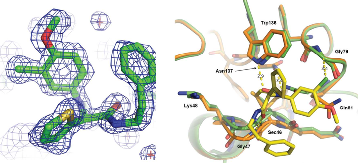
GPX4 consists of a compact monomer whose catalytic activity depends on a redox-active selenocysteine residue. Studies have indicated that this catalytic core is adapted to directly interact with lipid hydroperoxides, which helps explain why it is well suited for membrane-proximal antioxidant repair (Sakamoto et al., 2017; Ursini et al., 2022). Additionally, high-resolution structures demonstrated the wild-type human GPX4 and its complex with the covalent inhibitor ML162, providing a direct basis for rational inhibitor design and for understanding its limited conventional binding pockets. Crystal structural analysis demonstrated that ML162 covalently binds Sec46 and occupies a shallow catalytic surface pocket involving key active-site residues, thereby clarifying the structural basis of GPX4 inhibition. As shown in Figure 1, the inhibitor engages the catalytic region without a deep classical binding pocket, which helps explain the long-standing challenge of designing highly drug-like GPX4 ligands (Moosmayer et al., 2021).
Recent studies have identified a fin-loop-like structure in GPX4 that supports membrane engagement and underlies neuroprotection from ferroptosis. Therefore, GPX4 can now be viewed not just as an antioxidant enzyme but also as a membrane-anchored repair factor that utilizes its spatial orientation to perform various biological functions (Lorenz et al., 2026; Ursini et al., 2022).
Figure 1. The structural basis of GPX4 catalysis and covalent inhibitor binding.
(Moosmayer et al., 2021)
3. Biosynthesis and regulation of GPX4
GPX4 is a selenoprotein, and its synthesis depends on specialized cellular processes required for selenocysteine incorporation; this biochemical dependency is important because selenium confers enzymatic advantages that protect it from overoxidation during peroxide detoxification (Ingold et al., 2018). The activity of GPX4 is also tightly linked to the system Xc-/GSH/GPX4 axis, in which glutathione serves as the reducing substrate for the conversion of phospholipid hydroperoxides into non-toxic lipid alcohols (Chen et al., 2024). Therefore, GPX4 activity can be disabled by inhibition or by depleting intracellular glutathione stores via impaired cystine uptake (Xie et al., 2023). GPX4 is not regulated in isolation but operates within a broad ferroptosis defense mechanism, including GPX4-independent systems such as coenzyme Q10 (CoQ10) reduction mediated by ferroptosis suppressor protein 1 (FSP1) and mitochondrial defense pathways.
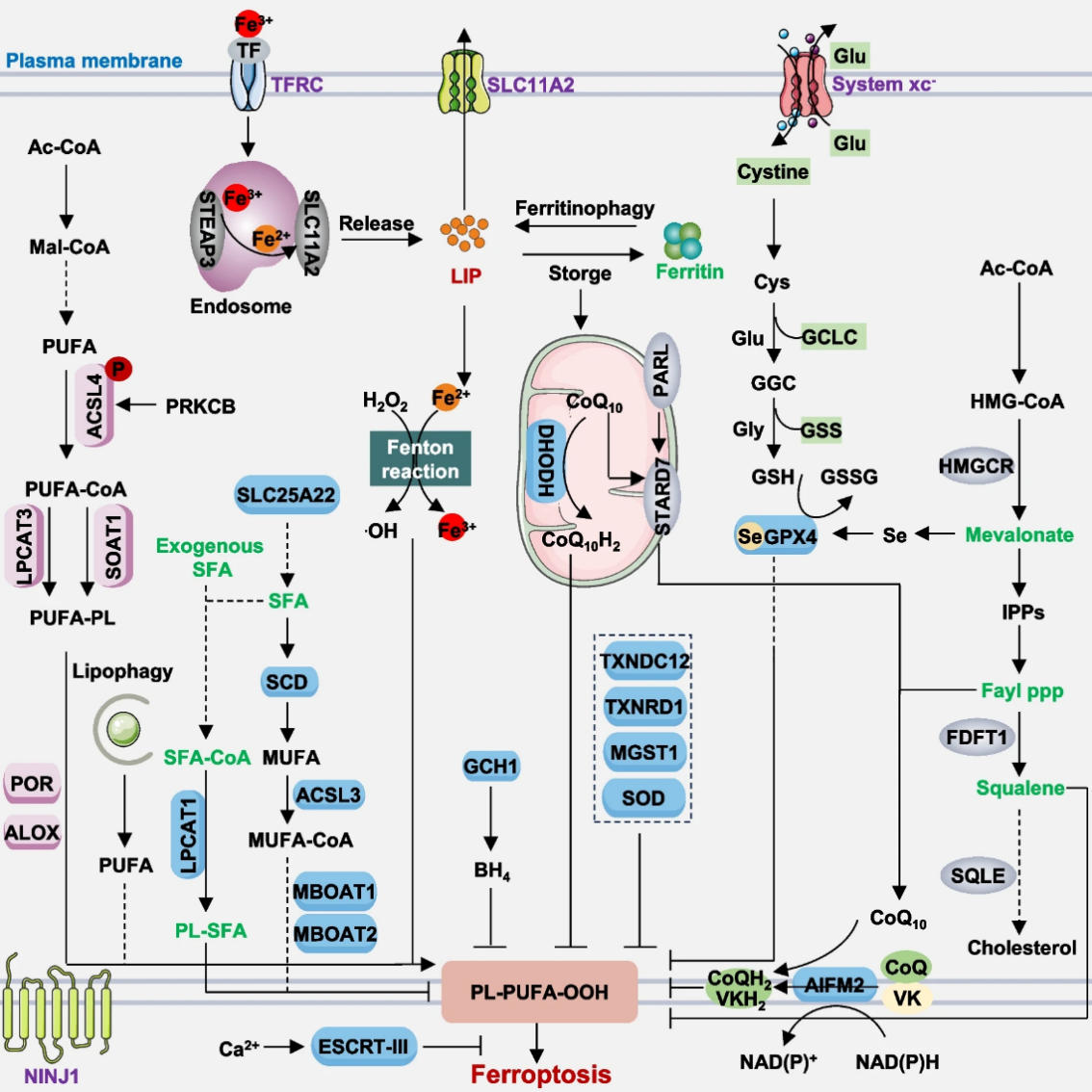
Ferroptosis arises from an imbalance between oxidant production and antioxidant defense, with iron accumulation, phospholipid peroxidation, and GPX4-dependent protection forming the core regulatory framework of the pathway. As summarized in Figure 2, both transporter-driven and enzyme-regulated routes converge on lipid peroxide accumulation and membrane injury, establishing the mechanistic context in which GPX4 suppresses ferroptosis (Chen et al., 2024). Studies have shown that GPX4 is a primary mechanism for suppressing phospholipid peroxide accumulation in most cell types, and its regulation occurs at multiple levels, including transcriptional control, post-transcriptional modulation, and context-specific stability/damage. These various levels have implications in normal tissue/tumor ferroptotic sensitivity (Qian et al., 2025; Zhang et al., 2024).
Figure 2. The core molecular mechanisms of ferroptosis and the GPX4-centered defense axis.
4. Biological functions of GPX4
A primary role of GPX4 is to eliminate phospholipid hydroperoxides and prevent further oxidation, thereby preserving membrane bilayers and structure, limiting oxidative chain reactions, and blocking the lethal phase of ferroptosis (Chen et al., 2024; Nishida Xavier Da Silva et al., 2022; Ursini et al., 2022). Membranes are essential for mitochondrial function, organelle signaling, and overall cell viability. GPX4 plays an integral role in multiple physiological processes beyond its role as a simple antioxidant enzyme. In addition, it has been linked to a critical role in maintaining structural integrity during development, supporting neurons, reproductive biology, and other autophagy-related cellular stress responses. Studies now describe it as a key molecule that connects lipid metabolism, redox balance, membrane biology, and regulated cell death, rather than as a terminal detoxifying enzyme (Xie et al., 2023; Zhang et al., 2024). Evidence that membrane association is important to GPX4's neuroprotective role supports a model in which it functions through both enzymatic catalysis and spatial control at vulnerable membrane surfaces (Lorenz et al., 2026).
5. GPX4 in disease
GPX4 has emerged as a major determinant of disease susceptibility because ferroptotic lipid damage contributes to numerous different pathological conditions. In cancer, numerous therapy-resistant, mesenchymal-like, or stress-adapted tumor cells utilize GPX4 to survive chronic oxidative stress, making GPX4 inhibition a promising anti-tumor therapeutic approach (Qian et al., 2025). Conversely, maintaining GPX4 activity in non-malignant tissues, such as the brain and heart, protects against injurious or inflammatory processes by limiting ferroptosis (Huang et al., 2024; Lorenz et al., 2026). The robustness of GPX4-mediated disease pathology is one of the most significant aspects of translating GPX4 biology into clinical practice. In oncology, reduced GPX4 activity may expose a therapeutic vulnerability. However, in other disorders, including cardiotoxicity, neurodegenerative diseases and ischemic injury, the same pathways may require reinforcement rather than suppression (Zhang et al., 2024). Several studies demonstrated the protective role of GPX4 by showing that pharmacological activation of GPX4 alleviated doxorubicin-induced cardiomyopathy, thereby supporting its therapeutic relevance in non-cancer disorders (Huang et al., 2024)
6. Therapeutic targeting of GPX4
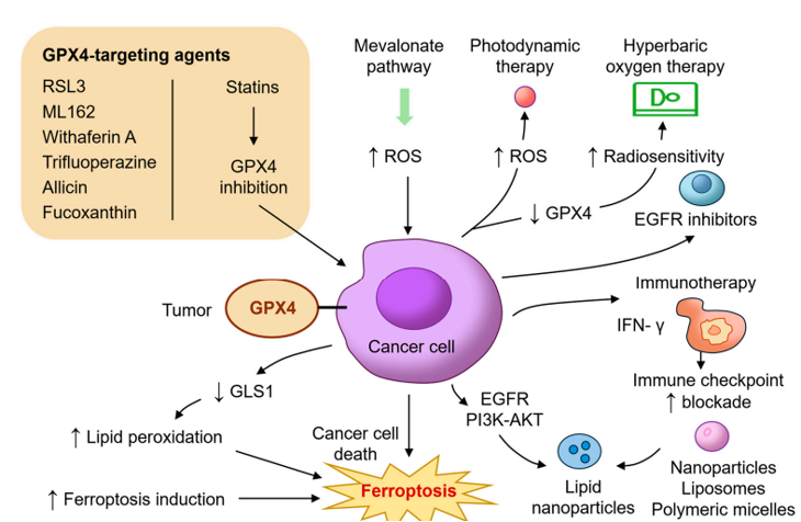
Therapeutic strategies targeting GPX4 now include covalent inhibitors, allosteric inhibitors, degraders, nanobody-based targeting systems, and GPX4-supporting compounds. Structural and medicinal chemistry studies have shown that direct targeting of GPX4 is challenging because the enzyme presents a relatively flat surface with limited conventional binding pockets (Moosmayer et al., 2021; Qian et al., 2025). This challenge has driven the development of non-classical targeting strategies, including peptide-derived inhibitors, covalent ligands and alternative binding modes (Sakamoto et al., 2017). One of the most recent advancements in GPX4 research was the creation of an anti-GPX4 nanobody, which demonstrated the ability to induce ferroptosis with improved selectivity and safety compared with classical small-molecule inhibitors. This advancement demonstrates the use of biological modalities to address intracellular targets (Li et al., 2024). Simultaneously, recent reviews on potential treatments that modulate GPX4 have highlighted that future significant advances will depend on greater selectivity, improved pharmacological control, and disease-specific deployment of either GPX4 inhibition or activation (Qian et al., 2025). Current therapeutic strategies targeting GPX4 include direct inhibitors, indirect modulators of upstream antioxidant pathways, and context-dependent approaches designed either to induce ferroptosis in resistant tumor cells or to preserve membrane integrity in non-malignant tissue injury. As summarized in Figure 3, GPX4-targeted intervention has emerged as a translational framework that links ferroptosis biology to disease-oriented therapeutic design (Lee et al., 2025).
Figure 3. Therapeutic implications of GPX4 targeting in ferroptosis-related disease contexts.
7. Methods used to study GPX4
Glutathione peroxidase 4 (GPX4) is a selenium-containing enzyme that occupies a central position in membrane redox biology as it directly reduces phospholipid hydroperoxides within biological membranes (Nishida Xavier Da Silva et al., 2022). While other glutathione peroxidases typically detoxify soluble hydroperoxides, GPX4 acts on complex lipid peroxides embedded in membranes and lipoproteins (Ursini et al., 2022). This biochemical specialization makes it a principal suppressor of ferroptosis, a regulated form of programmed cell death caused by iron-mediated lipid peroxidation and subsequent catastrophic damage to cellular membranes (Chen et al., 2024; Xie et al., 2023). The role of GPX4 extends beyond antioxidant defense. Recent studies have placed it at the center of several biological pathways that govern cancer cell survival, neurodegenerative diseases, inflammation-related tissue injuries, cardiac toxicity, metabolic syndrome and ischemic/reperfusion injuries (Huang et al., 2024; Zhang et al., 2024). The fact that phospholipid peroxidation within cellular membranes represents a common injury pathway in multiple disorders has led to GPX4 emerging as a key potential disease biomarker and therapeutic target in ferroptosis-centered drug development (Qian et al., 2025).
2. Structural characteristics of GPX4
GPX4 consists of a compact monomer whose catalytic activity depends on a redox-active selenocysteine residue. Studies have indicated that this catalytic core is adapted to directly interact with lipid hydroperoxides, which helps explain why it is well suited for membrane-proximal antioxidant repair (Sakamoto et al., 2017; Ursini et al., 2022). Additionally, high-resolution structures demonstrated the wild-type human GPX4 and its complex with the covalent inhibitor ML162, providing a direct basis for rational inhibitor design and for understanding its limited conventional binding pockets. Crystal structural analysis demonstrated that ML162 covalently binds Sec46 and occupies a shallow catalytic surface pocket involving key active-site residues, thereby clarifying the structural basis of GPX4 inhibition. As shown in Figure 1, the inhibitor engages the catalytic region without a deep classical binding pocket, which helps explain the long-standing challenge of designing highly drug-like GPX4 ligands (Moosmayer et al., 2021).
Recent studies have identified a fin-loop-like structure in GPX4 that supports membrane engagement and underlies neuroprotection from ferroptosis. Therefore, GPX4 can now be viewed not just as an antioxidant enzyme but also as a membrane-anchored repair factor that utilizes its spatial orientation to perform various biological functions (Lorenz et al., 2026; Ursini et al., 2022).
Figure 1. The structural basis of GPX4 catalysis and covalent inhibitor binding.
(Moosmayer et al., 2021)
3. Biosynthesis and regulation of GPX4
GPX4 is a selenoprotein, and its synthesis depends on specialized cellular processes required for selenocysteine incorporation; this biochemical dependency is important because selenium confers enzymatic advantages that protect it from overoxidation during peroxide detoxification (Ingold et al., 2018). The activity of GPX4 is also tightly linked to the system Xc-/GSH/GPX4 axis, in which glutathione serves as the reducing substrate for the conversion of phospholipid hydroperoxides into non-toxic lipid alcohols (Chen et al., 2024). Therefore, GPX4 activity can be disabled by inhibition or by depleting intracellular glutathione stores via impaired cystine uptake (Xie et al., 2023). GPX4 is not regulated in isolation but operates within a broad ferroptosis defense mechanism, including GPX4-independent systems such as coenzyme Q10 (CoQ10) reduction mediated by ferroptosis suppressor protein 1 (FSP1) and mitochondrial defense pathways.
Ferroptosis arises from an imbalance between oxidant production and antioxidant defense, with iron accumulation, phospholipid peroxidation, and GPX4-dependent protection forming the core regulatory framework of the pathway. As summarized in Figure 2, both transporter-driven and enzyme-regulated routes converge on lipid peroxide accumulation and membrane injury, establishing the mechanistic context in which GPX4 suppresses ferroptosis (Chen et al., 2024). Studies have shown that GPX4 is a primary mechanism for suppressing phospholipid peroxide accumulation in most cell types, and its regulation occurs at multiple levels, including transcriptional control, post-transcriptional modulation, and context-specific stability/damage. These various levels have implications in normal tissue/tumor ferroptotic sensitivity (Qian et al., 2025; Zhang et al., 2024).
Figure 2. The core molecular mechanisms of ferroptosis and the GPX4-centered defense axis.
4. Biological functions of GPX4
A primary role of GPX4 is to eliminate phospholipid hydroperoxides and prevent further oxidation, thereby preserving membrane bilayers and structure, limiting oxidative chain reactions, and blocking the lethal phase of ferroptosis (Chen et al., 2024; Nishida Xavier Da Silva et al., 2022; Ursini et al., 2022). Membranes are essential for mitochondrial function, organelle signaling, and overall cell viability. GPX4 plays an integral role in multiple physiological processes beyond its role as a simple antioxidant enzyme. In addition, it has been linked to a critical role in maintaining structural integrity during development, supporting neurons, reproductive biology, and other autophagy-related cellular stress responses. Studies now describe it as a key molecule that connects lipid metabolism, redox balance, membrane biology, and regulated cell death, rather than as a terminal detoxifying enzyme (Xie et al., 2023; Zhang et al., 2024). Evidence that membrane association is important to GPX4's neuroprotective role supports a model in which it functions through both enzymatic catalysis and spatial control at vulnerable membrane surfaces (Lorenz et al., 2026).
5. GPX4 in disease
GPX4 has emerged as a major determinant of disease susceptibility because ferroptotic lipid damage contributes to numerous different pathological conditions. In cancer, numerous therapy-resistant, mesenchymal-like, or stress-adapted tumor cells utilize GPX4 to survive chronic oxidative stress, making GPX4 inhibition a promising anti-tumor therapeutic approach (Qian et al., 2025). Conversely, maintaining GPX4 activity in non-malignant tissues, such as the brain and heart, protects against injurious or inflammatory processes by limiting ferroptosis (Huang et al., 2024; Lorenz et al., 2026). The robustness of GPX4-mediated disease pathology is one of the most significant aspects of translating GPX4 biology into clinical practice. In oncology, reduced GPX4 activity may expose a therapeutic vulnerability. However, in other disorders, including cardiotoxicity, neurodegenerative diseases and ischemic injury, the same pathways may require reinforcement rather than suppression (Zhang et al., 2024). Several studies demonstrated the protective role of GPX4 by showing that pharmacological activation of GPX4 alleviated doxorubicin-induced cardiomyopathy, thereby supporting its therapeutic relevance in non-cancer disorders (Huang et al., 2024)
6. Therapeutic targeting of GPX4
Therapeutic strategies targeting GPX4 now include covalent inhibitors, allosteric inhibitors, degraders, nanobody-based targeting systems, and GPX4-supporting compounds. Structural and medicinal chemistry studies have shown that direct targeting of GPX4 is challenging because the enzyme presents a relatively flat surface with limited conventional binding pockets (Moosmayer et al., 2021; Qian et al., 2025). This challenge has driven the development of non-classical targeting strategies, including peptide-derived inhibitors, covalent ligands and alternative binding modes (Sakamoto et al., 2017). One of the most recent advancements in GPX4 research was the creation of an anti-GPX4 nanobody, which demonstrated the ability to induce ferroptosis with improved selectivity and safety compared with classical small-molecule inhibitors. This advancement demonstrates the use of biological modalities to address intracellular targets (Li et al., 2024). Simultaneously, recent reviews on potential treatments that modulate GPX4 have highlighted that future significant advances will depend on greater selectivity, improved pharmacological control, and disease-specific deployment of either GPX4 inhibition or activation (Qian et al., 2025). Current therapeutic strategies targeting GPX4 include direct inhibitors, indirect modulators of upstream antioxidant pathways, and context-dependent approaches designed either to induce ferroptosis in resistant tumor cells or to preserve membrane integrity in non-malignant tissue injury. As summarized in Figure 3, GPX4-targeted intervention has emerged as a translational framework that links ferroptosis biology to disease-oriented therapeutic design (Lee et al., 2025).
Figure 3. Therapeutic implications of GPX4 targeting in ferroptosis-related disease contexts.
7. Methods used to study GPX4
GPX4 is studied using structural, biochemical and functional approaches. At the expression level, RT-qPCR, Western blotting, immunofluorescence, and immunohistochemistry are widely used to assess transcript abundance, protein localization, and tissue distribution. These approaches are commonly paired with measurements of glutathione status and lipid peroxidation to determine whether GPX4-associated ferroptosis has been engaged (Chen et al., 2024; Xie et al., 2023). Functional GPX4 studies typically require more than just expression data. Rescue experiments using ferroptosis inhibitors, direct lipid ROS assays, phospholipid oxidation profiling, and structure-based inhibitor studies are necessary to show whether GPX4 is mechanistically involved in a given phenotype (Li et al., 2024; Moosmayer et al., 2021). This combination of structural, biochemical, and cell-based evidence has become particularly important because ferroptosis intersects with several parallel redox systems and cannot be assigned confidently on morphology alone.
8. Conclusions
GPX4 is an important enzyme that protects membrane lipids from oxidative damage and helps prevent ferroptotic cell death. By reducing harmful phospholipid hydroperoxides, it supports cell survival, preserves membrane integrity, and contributes to normal tissue function. This role is relevant across several major disease areas, including cancer, neurodegeneration, cardiotoxicity, and inflammatory injury. Its biological importance extends beyond basic antioxidant defense, as it links lipid redox balance to disease development and treatment response. Therefore, GPX4 serves not only as a key regulator of cellular protection but also as a promising target for future therapeutic strategies.
References
8. Conclusions
GPX4 is an important enzyme that protects membrane lipids from oxidative damage and helps prevent ferroptotic cell death. By reducing harmful phospholipid hydroperoxides, it supports cell survival, preserves membrane integrity, and contributes to normal tissue function. This role is relevant across several major disease areas, including cancer, neurodegeneration, cardiotoxicity, and inflammatory injury. Its biological importance extends beyond basic antioxidant defense, as it links lipid redox balance to disease development and treatment response. Therefore, GPX4 serves not only as a key regulator of cellular protection but also as a promising target for future therapeutic strategies.
References
Chen, F., Kang, R., Tang, D., and Liu, J. (2024). Ferroptosis: principles and significance in health and disease. J. Hematol. Onco., 17, 41. https://doi.org/10.1186/s13045-024-01564-3
Huang, C., Guo, Y., Li, T., Sun, G., Yang, J., Wang, Y., Xiang, Y., Wang, L., Jin, M., Li, J., Zhou, Y., Han, B., Huang, R., Qiu, J., Tan, Y., Hu, J., Wei, Y., Wu, B., Mao, Y., Lei, L., Song, X., Li, S., Wang, Y., and Zhang, T. (2024). Pharmacological activation of GPX4 ameliorates doxorubicin-induced cardiomyopathy. Redox Biol. 70, 103024. https://doi.org/10.1016/j.redox.2023.103024
Ingold, I., Berndt, C., Schmitt, S., Doll, S., Poschmann, G., Buday, K., Roveri, A., Peng, X., Porto Freitas, F., Seibt, T., Mehr, L., Aichler, M., Walch, A., Lamp, D., Jastroch, M., Miyamoto, S., Wurst, W., Ursini, F., Arnér, E.S.J., Fradejas-Villar, N., Schweizer, U., Zischka, H., Friedmann Angeli, J.P., and Conrad, M. (2018). Selenium Utilization by GPX4 Is Required to Prevent Hydroperoxide-Induced Ferroptosis. Cell 172, 409-422.e21. https://doi.org/10.1016/j.cell.2017.11.048
Huang, C., Guo, Y., Li, T., Sun, G., Yang, J., Wang, Y., Xiang, Y., Wang, L., Jin, M., Li, J., Zhou, Y., Han, B., Huang, R., Qiu, J., Tan, Y., Hu, J., Wei, Y., Wu, B., Mao, Y., Lei, L., Song, X., Li, S., Wang, Y., and Zhang, T. (2024). Pharmacological activation of GPX4 ameliorates doxorubicin-induced cardiomyopathy. Redox Biol. 70, 103024. https://doi.org/10.1016/j.redox.2023.103024
Ingold, I., Berndt, C., Schmitt, S., Doll, S., Poschmann, G., Buday, K., Roveri, A., Peng, X., Porto Freitas, F., Seibt, T., Mehr, L., Aichler, M., Walch, A., Lamp, D., Jastroch, M., Miyamoto, S., Wurst, W., Ursini, F., Arnér, E.S.J., Fradejas-Villar, N., Schweizer, U., Zischka, H., Friedmann Angeli, J.P., and Conrad, M. (2018). Selenium Utilization by GPX4 Is Required to Prevent Hydroperoxide-Induced Ferroptosis. Cell 172, 409-422.e21. https://doi.org/10.1016/j.cell.2017.11.048
Lee, J., Seo, Y., and Roh, J.-L. (2025). Emerging Therapeutic Strategies Targeting GPX4-Mediated Ferroptosis in Head and Neck Cancer. Int. J. Mol. Sci. 26, 6452. https://doi.org/10.3390/ijms26136452
Li, X., Li, Y., Xie, A., Chen, F., Wang, J., Zhou, J., Xu, X., Xu, Z., Wang, Y., and Qiu, X. (2024). A potent and selective anti-glutathione peroxidase 4 nanobody as a ferroptosis inducer. Chem. Sci. 15, 19420–19431. https://doi.org/10.1039/D4SC05448B
Lorenz, S.M., Wahida, A., Bostock, M.J., Seibt, T., Santos Dias Mourão, A., Levkina, A., Trümbach, D., Soudy, M., Emler, D., Rothammer, N., Woo, M.S., Sonner, J.K., Novikova, M., Henkelmann, B., Aldrovandi, M., Kaemena, D.F., Mishima, E., Vermonden, P., Zong, Z., Chen, D., Nakamura, T., Ito, J., Doll, S., Proneth, B., Bürkle, E., Rizzollo, F., Escamilla Ayala, A., Napolitano, V., Kolonko-Adamska, M., Gaussmann, S., Merl-Pham, J., Hauck, S., Pertek, A., Orschmann, T., Van San, E., Vanden Berghe, T., Hass, D., Maida, A., Frenz, J.M., Pedrera, L., Dolga, A., Kraiger, M., Hrabé De Angelis, M., Fuchs, H., Ebert, G., Lenberg, J., Friedman, J., Scale, C., Agostinis, P., Zimprich, A., Vogt-Weisenhorn, D., Garrett, L., Hölter, S.M., Wurst, W., Glaab, E., Lewerenz, J., Popper, B., Sieben, C., Steinacker, P., Zischka, H., Garcia-Saez, A.J., Tietze, A., Ramesh, S.K., Ayton, S., Vincendeau, M., Friese, M.A., Wigby, K., Sattler, M., Mann, M., Ingold, I., Jayavelu, A.K., Popowicz, G.M., and Conrad, M. (2026). A fin-loop-like structure in GPX4 underlies neuroprotection from ferroptosis. Cell 189, 287-306.e35. https://doi.org/10.1016/j.cell.2025.11.014
Moosmayer, D., Hilpmann, A., Hoffmann, J., Schnirch, L., Zimmermann, K., Badock, V., Furst, L., Eaton, J.K., Viswanathan, V.S., Schreiber, S.L., Gradl, S., and Hillig, R.C. (2021). Crystal structures of the selenoprotein glutathione peroxidase 4 in its apo form and in complex with the covalently bound inhibitor ML162. Acta Crystallogr. Sect. Struct. Biol. 77, 237–248. https://doi.org/10.1107/S2059798320016125
Nishida Xavier Da Silva, T., Friedmann Angeli, J.P., and Ingold, I. (2022). GPX4: old lessons, new features. Biochem. Soc. Trans. 50, 1205–1213. https://doi.org/10.1042/BST20220682
Qian, J.-Y., Lou, C.-Y., Chen, Y.-L., Ma, L.-F., Hou, W., and Zhan, Z.-J. (2025). A prospective therapeutic strategy: GPX4-targeted ferroptosis mediators. Eur. J. Med. Chem. 281, 117015. https://doi.org/10.1016/j.ejmech.2024.117015
Sakamoto, K., Sogabe, S., Kamada, Y., Matsumoto, S., Kadotani, A., Sakamoto, J., and Tani, A. (2017). Discovery of GPX4 inhibitory peptides from random peptide T7 phage display and subsequent structural analysis. Biochem. Biophys. Res. Commun. 482, 195–201. https://doi.org/10.1016/j.bbrc.2016.11.035
Ursini, F., Bosello Travain, V., Cozza, G., Miotto, G., Roveri, A., Toppo, S., and Maiorino, M. (2022). A white paper on Phospholipid Hydroperoxide Glutathione Peroxidase (GPx4) forty years later. Free Radic. Biol. Med. 188, 117–133. https://doi.org/10.1016/j.freeradbiomed.2022.06.227
Xie, Y., Kang, R., Klionsky, D.J., and Tang, D. (2023). GPX4 in cell death, autophagy, and disease. Autophagy 19, 2621–2638. https://doi.org/10.1080/15548627.2023.2218764
Zhang, W., Liu, Y., Liao, Y., Zhu, C., and Zou, Z. (2024). GPX4, ferroptosis, and diseases. Biomed. Pharmacother. 174, 116512. https://doi.org/10.1016/j.biopha.2024.116512
Lorenz, S.M., Wahida, A., Bostock, M.J., Seibt, T., Santos Dias Mourão, A., Levkina, A., Trümbach, D., Soudy, M., Emler, D., Rothammer, N., Woo, M.S., Sonner, J.K., Novikova, M., Henkelmann, B., Aldrovandi, M., Kaemena, D.F., Mishima, E., Vermonden, P., Zong, Z., Chen, D., Nakamura, T., Ito, J., Doll, S., Proneth, B., Bürkle, E., Rizzollo, F., Escamilla Ayala, A., Napolitano, V., Kolonko-Adamska, M., Gaussmann, S., Merl-Pham, J., Hauck, S., Pertek, A., Orschmann, T., Van San, E., Vanden Berghe, T., Hass, D., Maida, A., Frenz, J.M., Pedrera, L., Dolga, A., Kraiger, M., Hrabé De Angelis, M., Fuchs, H., Ebert, G., Lenberg, J., Friedman, J., Scale, C., Agostinis, P., Zimprich, A., Vogt-Weisenhorn, D., Garrett, L., Hölter, S.M., Wurst, W., Glaab, E., Lewerenz, J., Popper, B., Sieben, C., Steinacker, P., Zischka, H., Garcia-Saez, A.J., Tietze, A., Ramesh, S.K., Ayton, S., Vincendeau, M., Friese, M.A., Wigby, K., Sattler, M., Mann, M., Ingold, I., Jayavelu, A.K., Popowicz, G.M., and Conrad, M. (2026). A fin-loop-like structure in GPX4 underlies neuroprotection from ferroptosis. Cell 189, 287-306.e35. https://doi.org/10.1016/j.cell.2025.11.014
Moosmayer, D., Hilpmann, A., Hoffmann, J., Schnirch, L., Zimmermann, K., Badock, V., Furst, L., Eaton, J.K., Viswanathan, V.S., Schreiber, S.L., Gradl, S., and Hillig, R.C. (2021). Crystal structures of the selenoprotein glutathione peroxidase 4 in its apo form and in complex with the covalently bound inhibitor ML162. Acta Crystallogr. Sect. Struct. Biol. 77, 237–248. https://doi.org/10.1107/S2059798320016125
Nishida Xavier Da Silva, T., Friedmann Angeli, J.P., and Ingold, I. (2022). GPX4: old lessons, new features. Biochem. Soc. Trans. 50, 1205–1213. https://doi.org/10.1042/BST20220682
Qian, J.-Y., Lou, C.-Y., Chen, Y.-L., Ma, L.-F., Hou, W., and Zhan, Z.-J. (2025). A prospective therapeutic strategy: GPX4-targeted ferroptosis mediators. Eur. J. Med. Chem. 281, 117015. https://doi.org/10.1016/j.ejmech.2024.117015
Sakamoto, K., Sogabe, S., Kamada, Y., Matsumoto, S., Kadotani, A., Sakamoto, J., and Tani, A. (2017). Discovery of GPX4 inhibitory peptides from random peptide T7 phage display and subsequent structural analysis. Biochem. Biophys. Res. Commun. 482, 195–201. https://doi.org/10.1016/j.bbrc.2016.11.035
Ursini, F., Bosello Travain, V., Cozza, G., Miotto, G., Roveri, A., Toppo, S., and Maiorino, M. (2022). A white paper on Phospholipid Hydroperoxide Glutathione Peroxidase (GPx4) forty years later. Free Radic. Biol. Med. 188, 117–133. https://doi.org/10.1016/j.freeradbiomed.2022.06.227
Xie, Y., Kang, R., Klionsky, D.J., and Tang, D. (2023). GPX4 in cell death, autophagy, and disease. Autophagy 19, 2621–2638. https://doi.org/10.1080/15548627.2023.2218764
Zhang, W., Liu, Y., Liao, Y., Zhu, C., and Zou, Z. (2024). GPX4, ferroptosis, and diseases. Biomed. Pharmacother. 174, 116512. https://doi.org/10.1016/j.biopha.2024.116512


