


HMGB1: A Redox-Regulated Switch in Cell Death and Inflammation
1. Introduction
High mobility group box 1 (HMGB1) is a highly conserved, widely expressed non-histone chromatin protein that regulates cell survival and death based on its subcellular location and redox state (Tang et al., 2023). In the nucleus, it acts as an architectural DNA chaperone that facilitates nucleosome remodeling, transcription, replication, and DNA repair (Kwak et al., 2026). During cellular stress, infection, or death, it undergoes translocation and is released into the extracellular space, functioning as a damage-associated molecular pattern (DAMP) that engages pattern-recognition receptors and initiates innate immune activation. This multi-functional property, including its structure, distinguishes it from several chromatin-associated proteins and underlies its involvement in a wide range of pathological processes (Chen et al., 2022; Kwak et al., 2026).
HMGB1 was initially isolated from calf thymus chromatin and characterized as a structural component of chromatin (Tang et al., 2023). Its immunological significance was established after extracellular HMGB1 was released from necrotic and stressed cells, alerting the innate immune system, analogous to other DAMPs (Chen et al., 2022). Previous studies have associated HMGB1 with sepsis, ischemic injury, autoimmune and autoinflammatory skin disease, neurodegeneration, and numerous malignancies, and have shown that it can protect against these defects, depending on tissue, redox state, and receptor engagement (Fan et al., 2025; Lv et al., 2025).
2. Structural Architecture and Redox Isoforms
HMGB1 is a 215-amino-acid, approximately 25-kDa protein composed of two tandem HMG-box domains, designated box A and box B, followed by an acidic carboxy-terminal tail enriched in glutamate and aspartate residues (Yuan et al., 2020). Each HMG box adopts an L-shaped fold comprising three α-helices, a conformation conserved between the two domains yet functionally separated. Box A is associated mainly with anti-inflammatory properties and has been developed as a competitive antagonist of full-length HMGB1, whereas Box B contains the segment required for TLR4 engagement and is principally responsible for the protein’s pro-inflammatory signaling capacity (Shen et al., 2024). The acidic C-terminal tail modulates the DNA-binding affinity of the two boxes through an intramolecular interaction and additionally contains a short antimicrobial peptide sequence, indicating that HMGB1 possesses functional capacities beyond chromatin architecture and immune signaling (Tang et al., 2023).
The function of HMGB1 is significantly governed by the redox status of three cysteine residues located at positions 23, 45, and 106. In the fully reduced state, all three cysteines retain free thiol groups, and the protein exhibits chemoattractant activity, recruiting immune cells independently of classical cytokine signaling. Oxidative conditions promote the formation of an intramolecular disulfide bond between Cys23 and Cys45, generating disulfide HMGB1, the isoform required for MD-2/TLR4 engagement and NF-κB-dependent cytokine induction. Further oxidation converts all cysteines to sulfonate derivatives, leading to a fully oxidized isoform that has lost both DNA-binding capacity and receptor affinity. A fourth isoform, generated by intermolecular disulfide bond formation between Cys106 residues of two HMGB1 molecules, produces two species with markedly greater affinity for TLR2 and TLR4 than a single protein (Kwak et al., 2026). These four redox isoforms share the same polypeptide sequence but exhibit distinct receptor specificities and biological activities, establishing redox state as a primary determinant of HMGB1 function.
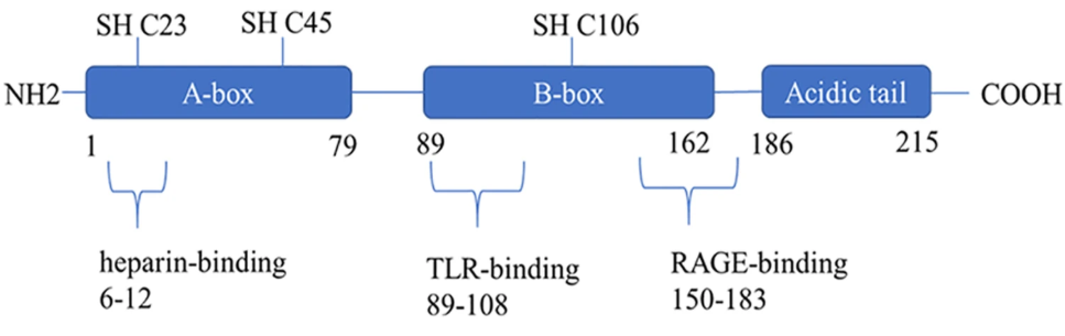
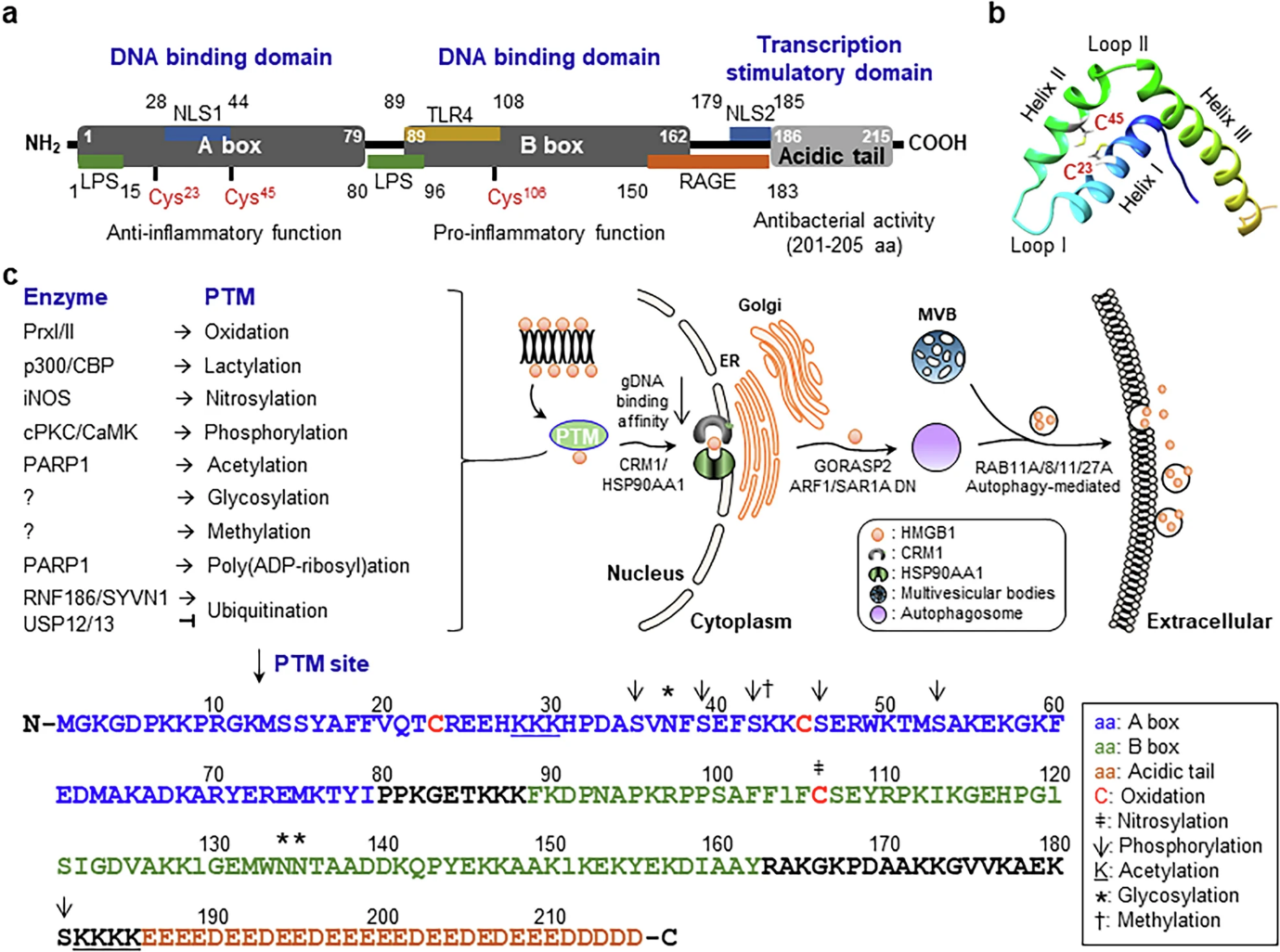
Figure 1. (A) Schematic representation of HMGB1 domain architecture, including the A-box and B-box domains, the acidic C-terminal tail, the two nuclear localization signals, and the binding regions for lipopolysaccharide, TLR4, and RAGE. (B) The redox isoforms of HMGB1, including the dimeric (Di-HMGB1) species formed via intermolecular Cys106 disulfide bonding and its enhanced receptor affinity relative to the monomer.
1. Biosynthesis, Post-Translational Regulation and Release of HMGB1
HMGB1 biosynthesis follows the conventional pathway of transcription, translation, and nuclear import mediated by two nuclear localization signals; however, its subcellular distribution and extracellular release are regulated predominantly by post-translational modifications rather than by alterations in expression levels (Tang et al., 2023). Its acetylation by the histone acetyltransferases p300/CBP reduces its chromatin affinity and promotes cytoplasm translocation. Similarly, phosphorylation by classical protein kinase C and by calcium/calmodulin-dependent kinase IV facilitates nucleo-cytoplasmic shuttling, while lactylation, driven by elevated intracellular lactate under inflammatory conditions, promotes exosomal HMGB1 release during sepsis (Kwak et al., 2026). Cytoplasmic HMGB1 is then incorporated into secretory lysosomes and autophagosomes through a pathway that involves ATG5, ATG7, and components of the Golgi and vesicular transport machinery, enabling active secretion that can lead to the disruption of the plasma membrane (Chen et al., 2022).
The mode of cell death determines both the timing and the redox state of HMGB1 release, with direct consequences for its subsequent immunological activity. Necrotic cell death releases the reduced isoform during the initial stages, with oxidation occurring as the process continues. Pyroptosis, mediated by inflammasome activation and gasdermin-dependent pore formation, releases the disulfide isoform mainly alongside interleukin-1β and interleukin-18. Apoptosis produces little immune activation. HMGB1 remains trapped within apoptotic bodies during this process, and any small amount that escapes is already heavily oxidized, thereby losing the ability to trigger an immune response (Kwak et al., 2026). Ferroptosis represents an intermediate between these two extremes and is recently understood. During ferroptosis, the accumulation of lipid hydroperoxides damages the nuclear membrane before the plasma membrane breaks down, allowing HMGB1 to enter the cytoplasm earlier than it would during classical necrosis. Autophagy-related proteins then help carry it out of the cell (Lv et al., 2025). The temporal stage at which HMGB1 is released during ferroptosis highly influences its immunological effects: early-stage release promotes cell maturation and antitumor immunity, while late-stage release, associated with membrane failure, is linked with reduced immunity, indicating that HMGB1 release should be regarded as a graded process rather than a binary event (Chen et al., 2022).
2. Physiological Roles
HMGB1 functions primarily as an architectural protein that binds the minor groove of DNA, induces bending, and facilitates nucleosome remodeling, transcriptional regulation, and DNA repair (Tang et al., 2023). In the cytoplasm, the reduced HMGB1 directly binds Beclin-1, displacing Bcl-2 and promoting autophagosome formation; this interaction supports mitophagy and broader mitochondrial quality control (Chen et al., 2022). In addition to its DNA-binding activity, HMGB1 is reported to exhibit chaperone-like activity and suppress the aggregation of polyglutamine repeats implicated in neurodegenerative disease, a function that extends the protein’s role beyond chromatin regulation and immune signaling into general proteostatic maintenance. In the extracellular compartment, reduced HMGB1 forms a heterocomplex with the chemokine CXCL12, which signals through CXCR4 to recruit immune and stem cells to sites of tissue injury, thereby contributing to repair rather than inflammatory responses (Kwak et al., 2026). A related but distinct activity has been found for HMGB1 released by natural killer cells, which can directly kill colorectal cancer cells by binding to and blocking an enzyme called pyruvate kinase M2. This forces cancer cells to rely solely on glycolysis for energy, and it occurs without involving TLR4 or RAGE signaling (Gdynia et al., 2016). These findings collectively indicate that HMGB1 contributes to tissue repair, immune cell recruitment, regulation of autophagy, and direct antitumor cytotoxicity through mechanisms independent of its canonical pro-inflammatory signaling.
5. HMGB1 in Disease
5.1 Sepsis and Systemic Inflammation
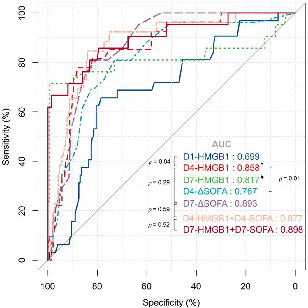
HMGB1 was first identified as a late-acting driver of death in mouse models of sepsis, and clinical studies have confirmed its relevance to sepsis in humans. Serum HMGB1 levels rise with sepsis severity and closely track organ damage. In one cohort study, HMGB1 predicted patient outcomes about as well as other established sepsis biomarkers, with an area under the receiver operating characteristic curve (AUC) of approximately 0.78 (Jiang, 2024). A recent study of 232 sepsis patients found that HMGB1 measured just 4 days after admission predicted 28-day mortality as accurately as a dynamic organ-failure score, which normally takes a full week. This suggests that a single, well-timed HMGB1 measurement could identify high-risk patients well before conventional scoring methods (Su et al., 2026).
(Su et al., 2026)
Figure 2. Receiver operating characteristic curves comparing serum HMGB1 concentration against the SOFA score across multiple measurement time points for prediction of 28-day mortality in sepsis.
5.2 Ferroptosis-Associated Disease across Organ Systems
The relationship between HMGB1 and ferroptosis has emerged as one of the most rapidly increasing areas of focus in HMGB1 research. Some studies traced the HMGB1-ferroptosis connection across nearly every major organ system, including acute lung injury and pulmonary hypertension in the lungs, drug-induced liver injury, hepatic fibrosis, and hepatocellular carcinoma in the liver. Additionally, ischemic stroke, traumatic brain injury, and spinal cord injury in the nervous system, ischemia-reperfusion injury and drug-induced cardiomyopathy in the heart, and acute and chronic kidney injury in the kidneys (Lv et al., 2025). The underlying mechanism was consistent across these tissues: HMGB1 released from ferroptotic cells activated RAGE and TLR4 on nearby macrophages and epithelial cells, thereby worsening iron buildup and lipid peroxidation in these cells. This creates a self-reinforcing cycle of cell death and inflammation (Du et al., 2026).
5.3 Cancer
HMGB1 exhibits multiple activities in oncology. During chemotherapy- or radiotherapy-induced cell death, it is released from dying tumor cells, engages TLR4 on dendritic cells, and contributes to immunogenic cell death, a mechanism exploited therapeutically to enhance antitumor immune responses (Alhasan et al., 2025). Conversely, HMGB1 retained within surviving tumor cells enhances chromatin accessibility for DNA repair machinery following genotoxic stress, sustains pro-survival autophagy, and upregulates multidrug resistance transporters, collectively conferring resistance to subsequent therapeutic challenge (Shen et al., 2024). This multifunctional property has been well documented in hematologic malignancies, in which HMGB1 contributed to bone marrow microenvironmental signaling, chemoresistance, and inflammation-driven maintenance of leukemic stem cells across multiple blood cancer subtypes, thereby establishing it as a pivotal regulator in this disease category (Yuan et al., 2020).
5.4 Autoimmune, Dermatology and Central Nervous System Disease
Beyond oncology, HMGB1 also serves as both a biomarker and a contributing mechanism in autoimmune and autoinflammatory conditions, including psoriasis, vitiligo, atopic dermatitis, and alopecia areata, acting mainly through TLR2/TLR4/RAGE receptors (Fan et al., 2025). In the central nervous system, it acts as a shared driver behind several conditions, including ischemic and hemorrhagic stroke, traumatic brain injury, multiple sclerosis, and neurodegenerative disease. Its levels in blood and cerebrospinal fluid track closely with disease severity, making it a useful prognostic marker (Du et al., 2026). The consistency of this receptor-driven mechanism across different organ systems highlights HMGB1’s role as a broadly acting inflammatory mediator and helps explain the growing pharmaceutical interest in its target.
(Mao et al., 2022)
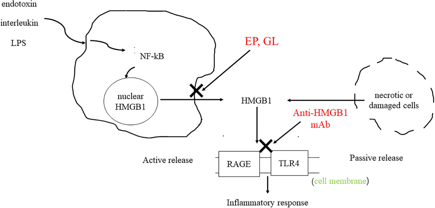
Figure 3. HMGB1 active and passive release mechanisms and downstream signaling via RAGE and TLR4 to drive pro-inflammatory cytokine production, with glycyrrhizin, ethyl pyruvate, and an anti-HMGB1 monoclonal antibody identified as potential pharmacological intervention points.
6. Therapeutic Targeting of HMGB1
The dependency of HMGB1’s harmful effects on its redox state and on a fairly small set of receptor-binding sites has made it an appealing drug target. Glycyrrhizin, a plant-derived compound that binds HMGB1 directly, is the recently best-studied inhibitor. It has shown benefits in models of acute liver failure, ischemic stroke, and ferroptosis-driven kidney injury, mainly by blocking the RAGE/TLR4-NF-κB signaling pathway (Lv et al., 2025). More recent drug-design studies have mapped the structural pockets on HMGB1 and its interactions with RAGE and TLR4. This helps researchers design small molecules and larger inhibitors that target specific protein-protein interactions, rather than suppressing HMGB1 (Shen et al., 2024).
Therapeutic targeting of HMGB1 in oncology is complicated by the protein’s contribution to immunogenic cell death during chemotherapy; indiscriminate inhibition risks attenuating a beneficial antitumor signal while amplifying a pathogenic one. Consequently, recent strategies have focused on selectively disrupting HMGB1’s intracellular pro-survival functions, including DNA repair support, pro-survival autophagy, and upregulation of drug efflux transporters, while preserving its extracellular immunostimulatory function (Alhasan et al., 2025). This compartment-specific approach helps explain why HMGB1-targeted treatments work well in inflammatory disease but have had mixed results in cancer.
7. Detection and Quantification of HMGB1
Enzyme-linked immunosorbent assay performed on serum or plasma remains the predominant method for clinical quantification of HMGB1 and is adequate for tracking total protein concentration across patient cohorts. However, this approach does not distinguish between redox isoforms, a limitation of considerable biological significance given the isoform-dependent receptor specificity (Kwak et al., 2026). Total serum HMGB1 concentration alone has proven to sufficiently track sepsis severity and mortality risk, despite the absence of isoform-resolved data (Jiang, 2024; Su et al., 2026). Isoform-specific quantification requires either redox-state-specific antibodies that discriminate among reduced, disulfide, and fully oxidized HMGB1, or mass spectrometry-based approaches that directly resolve the relevant cysteine modifications.
Western blotting and immunohistochemistry remain the standard tools for determining the localization of HMGB1 in cells or tissues, rather than measuring its circulating levels. These methods can show whether HMGB1 stays in the nucleus or has moved into the cytoplasm in a biopsy sample, and cytoplasmic HMGB1 has been linked to more aggressive cancers in several studies (Yuan et al., 2020). As redox-isoform-specific reagents become more widely available, clinical testing is likely to move toward measuring specific isoforms rather than just total HMGB1.
8. Conclusions
HMGB1 is a multifunctional, redox-regulated alarmin that connects chromatin architecture, regulated cell death, and innate immune signaling. Its biological activity is governed less by total abundance than by subcellular localization and the oxidation state of three critical cysteine residues. This allows the same protein to function as a chromatin chaperone in the resting nucleus, a chemoattractant in early stress, a potent TLR4/RAGE agonist once oxidized to its disulfide form, and an immunologically inert species upon full oxidation. This redox-dependent switch underlies its involvement in sepsis, ferroptosis-associated organ injury, autoimmune dermatologic disease, central nervous system disorders, and cancer, in which it can act as either a beneficial mediator of tissue repair and immunogenic cell death or a driver of chronic inflammation and therapy resistance, depending on cellular context and receptor engagement. The value of HMGB1 as a biomarker and therapeutic target is therefore closely tied to isoform- and compartment-specific resolution. Its growing relevance in inflammatory, oncology, and neurological disease makes it a strong candidate for biomarker development and compartment-selective therapeutic targeting.
References
Alhasan, B.A., Margulis, B.A., and Guzhova, I.V. (2025). HMGB1: A Central Node in Cancer Therapy Resistance. Int. J. Mol. Sci. 26, 12010. https://doi.org/10.3390/ijms262412010
Chen, R., Kang, R., and Tang, D. (2022). The mechanism of HMGB1 secretion and release. Exp. Mol. Med. 54, 91–102. https://doi.org/10.1038/s12276-022-00736-w
Du, O., Wu, Y.-J., Li, M.-Y., and Du, J.-R. (2026). The role of HMGB1 in central nervous system (CNS) diseases: mechanisms and therapeutic perspectives. Cytokine 198, 157099. https://doi.org/10.1016/j.cyto.2025.157099
Fan, J., He, K., Zhang, Y., Li, R., Yi, X., and Li, S. (2025). HMGB1: new biomarker and therapeutic target of autoimmune and autoinflammatory skin diseases. Front. Immunol. 16, 1569632. https://doi.org/10.3389/fimmu.2025.1569632
Gdynia, G., Sauer, S.W., Kopitz, J., Fuchs, D., Duglova, K., Ruppert, T., Miller, M., Pahl, J., Cerwenka, A., Enders, M., Mairbäurl, H., Kamiński, M.M., Penzel, R., Zhang, C., Fuller, J.C., Wade, R.C., Benner, A., Chang-Claude, J., Brenner, H., Hoffmeister, M., Zentgraf, H., Schirmacher, P., and Roth, W. (2016). The HMGB1 protein induces a metabolic type of tumour cell death by blocking aerobic respiration. Nat. Commun. 7, 10764. https://doi.org/10.1038/ncomms10764
Jiang, S. (2024). Correlation of serum H-FABP, sTREM-1, and HMGB1 levels with severity and prognosis of sepsis. Am. J. Transl. Res. 16, 5846–5855. https://doi.org/10.62347/KELZ4296
Kwak, M.S., Jung, S.F., Park, I.H., and Shin, J.-S. (2026). The redox-sensitive protein HMGB1: intracellular and extracellular roles. Exp. Mol. Med. 58, 345–356. https://doi.org/10.1038/s12276-026-01640-3
Lv, L., Wang, Y., Lv, X., and Miao, Q. (2025). Involvement of HMGB1-mediated ferroptosis in systemic diseases. Front. Cell Dev. Biol. 13, 1676941. https://doi.org/10.3389/fcell.2025.1676941
Mao, D., Zheng, Y., Xu, F., Han, X., and Zhao, H. (2022). HMGB1 in nervous system diseases: A common biomarker and potential therapeutic target. Front. Neurol. 13, 1029891. https://doi.org/10.3389/fneur.2022.1029891
Shen, P., Zhang, L., Jiang, X., Yu, B., and Zhang, J. (2024). Targeting HMGB1 and Its Interaction with Receptors: Challenges and Future Directions. J. Med. Chem. 67, 21671–21694. https://doi.org/10.1021/acs.jmedchem.4c01912
Su, Q., Yu, J., He, S., Fan, C., Chen, Y., Zhou, W., and Qiao, M. (2026). Serial assessment of serum HMGB1 and ΔSOFA for predicting 28-day mortality in sepsis patients: a prospective cohort study in the emergency department. BMC Emerg. Med. 26, 127. https://doi.org/10.1186/s12873-026-01547-2
Tang, D., Kang, R., Zeh, H.J., and Lotze, M.T. (2023). The multifunctional protein HMGB1: 50 years of discovery. Nat. Rev. Immunol. 23, 824–841. https://doi.org/10.1038/s41577-023-00894-6
Yuan, S., Liu, Z., Xu, Z., Liu, J., and Zhang, J. (2020). High mobility group box 1 (HMGB1): a pivotal regulator of hematopoietic malignancies. J. Hematol. Oncol. 13, 91. https://doi.org/10.1186/s13045-020-00920-3
High mobility group box 1 (HMGB1) is a highly conserved, widely expressed non-histone chromatin protein that regulates cell survival and death based on its subcellular location and redox state (Tang et al., 2023). In the nucleus, it acts as an architectural DNA chaperone that facilitates nucleosome remodeling, transcription, replication, and DNA repair (Kwak et al., 2026). During cellular stress, infection, or death, it undergoes translocation and is released into the extracellular space, functioning as a damage-associated molecular pattern (DAMP) that engages pattern-recognition receptors and initiates innate immune activation. This multi-functional property, including its structure, distinguishes it from several chromatin-associated proteins and underlies its involvement in a wide range of pathological processes (Chen et al., 2022; Kwak et al., 2026).
HMGB1 was initially isolated from calf thymus chromatin and characterized as a structural component of chromatin (Tang et al., 2023). Its immunological significance was established after extracellular HMGB1 was released from necrotic and stressed cells, alerting the innate immune system, analogous to other DAMPs (Chen et al., 2022). Previous studies have associated HMGB1 with sepsis, ischemic injury, autoimmune and autoinflammatory skin disease, neurodegeneration, and numerous malignancies, and have shown that it can protect against these defects, depending on tissue, redox state, and receptor engagement (Fan et al., 2025; Lv et al., 2025).
2. Structural Architecture and Redox Isoforms
HMGB1 is a 215-amino-acid, approximately 25-kDa protein composed of two tandem HMG-box domains, designated box A and box B, followed by an acidic carboxy-terminal tail enriched in glutamate and aspartate residues (Yuan et al., 2020). Each HMG box adopts an L-shaped fold comprising three α-helices, a conformation conserved between the two domains yet functionally separated. Box A is associated mainly with anti-inflammatory properties and has been developed as a competitive antagonist of full-length HMGB1, whereas Box B contains the segment required for TLR4 engagement and is principally responsible for the protein’s pro-inflammatory signaling capacity (Shen et al., 2024). The acidic C-terminal tail modulates the DNA-binding affinity of the two boxes through an intramolecular interaction and additionally contains a short antimicrobial peptide sequence, indicating that HMGB1 possesses functional capacities beyond chromatin architecture and immune signaling (Tang et al., 2023).
The function of HMGB1 is significantly governed by the redox status of three cysteine residues located at positions 23, 45, and 106. In the fully reduced state, all three cysteines retain free thiol groups, and the protein exhibits chemoattractant activity, recruiting immune cells independently of classical cytokine signaling. Oxidative conditions promote the formation of an intramolecular disulfide bond between Cys23 and Cys45, generating disulfide HMGB1, the isoform required for MD-2/TLR4 engagement and NF-κB-dependent cytokine induction. Further oxidation converts all cysteines to sulfonate derivatives, leading to a fully oxidized isoform that has lost both DNA-binding capacity and receptor affinity. A fourth isoform, generated by intermolecular disulfide bond formation between Cys106 residues of two HMGB1 molecules, produces two species with markedly greater affinity for TLR2 and TLR4 than a single protein (Kwak et al., 2026). These four redox isoforms share the same polypeptide sequence but exhibit distinct receptor specificities and biological activities, establishing redox state as a primary determinant of HMGB1 function.
Figure 1. (A) Schematic representation of HMGB1 domain architecture, including the A-box and B-box domains, the acidic C-terminal tail, the two nuclear localization signals, and the binding regions for lipopolysaccharide, TLR4, and RAGE. (B) The redox isoforms of HMGB1, including the dimeric (Di-HMGB1) species formed via intermolecular Cys106 disulfide bonding and its enhanced receptor affinity relative to the monomer.
1. Biosynthesis, Post-Translational Regulation and Release of HMGB1
HMGB1 biosynthesis follows the conventional pathway of transcription, translation, and nuclear import mediated by two nuclear localization signals; however, its subcellular distribution and extracellular release are regulated predominantly by post-translational modifications rather than by alterations in expression levels (Tang et al., 2023). Its acetylation by the histone acetyltransferases p300/CBP reduces its chromatin affinity and promotes cytoplasm translocation. Similarly, phosphorylation by classical protein kinase C and by calcium/calmodulin-dependent kinase IV facilitates nucleo-cytoplasmic shuttling, while lactylation, driven by elevated intracellular lactate under inflammatory conditions, promotes exosomal HMGB1 release during sepsis (Kwak et al., 2026). Cytoplasmic HMGB1 is then incorporated into secretory lysosomes and autophagosomes through a pathway that involves ATG5, ATG7, and components of the Golgi and vesicular transport machinery, enabling active secretion that can lead to the disruption of the plasma membrane (Chen et al., 2022).
The mode of cell death determines both the timing and the redox state of HMGB1 release, with direct consequences for its subsequent immunological activity. Necrotic cell death releases the reduced isoform during the initial stages, with oxidation occurring as the process continues. Pyroptosis, mediated by inflammasome activation and gasdermin-dependent pore formation, releases the disulfide isoform mainly alongside interleukin-1β and interleukin-18. Apoptosis produces little immune activation. HMGB1 remains trapped within apoptotic bodies during this process, and any small amount that escapes is already heavily oxidized, thereby losing the ability to trigger an immune response (Kwak et al., 2026). Ferroptosis represents an intermediate between these two extremes and is recently understood. During ferroptosis, the accumulation of lipid hydroperoxides damages the nuclear membrane before the plasma membrane breaks down, allowing HMGB1 to enter the cytoplasm earlier than it would during classical necrosis. Autophagy-related proteins then help carry it out of the cell (Lv et al., 2025). The temporal stage at which HMGB1 is released during ferroptosis highly influences its immunological effects: early-stage release promotes cell maturation and antitumor immunity, while late-stage release, associated with membrane failure, is linked with reduced immunity, indicating that HMGB1 release should be regarded as a graded process rather than a binary event (Chen et al., 2022).
2. Physiological Roles
HMGB1 functions primarily as an architectural protein that binds the minor groove of DNA, induces bending, and facilitates nucleosome remodeling, transcriptional regulation, and DNA repair (Tang et al., 2023). In the cytoplasm, the reduced HMGB1 directly binds Beclin-1, displacing Bcl-2 and promoting autophagosome formation; this interaction supports mitophagy and broader mitochondrial quality control (Chen et al., 2022). In addition to its DNA-binding activity, HMGB1 is reported to exhibit chaperone-like activity and suppress the aggregation of polyglutamine repeats implicated in neurodegenerative disease, a function that extends the protein’s role beyond chromatin regulation and immune signaling into general proteostatic maintenance. In the extracellular compartment, reduced HMGB1 forms a heterocomplex with the chemokine CXCL12, which signals through CXCR4 to recruit immune and stem cells to sites of tissue injury, thereby contributing to repair rather than inflammatory responses (Kwak et al., 2026). A related but distinct activity has been found for HMGB1 released by natural killer cells, which can directly kill colorectal cancer cells by binding to and blocking an enzyme called pyruvate kinase M2. This forces cancer cells to rely solely on glycolysis for energy, and it occurs without involving TLR4 or RAGE signaling (Gdynia et al., 2016). These findings collectively indicate that HMGB1 contributes to tissue repair, immune cell recruitment, regulation of autophagy, and direct antitumor cytotoxicity through mechanisms independent of its canonical pro-inflammatory signaling.
5. HMGB1 in Disease
5.1 Sepsis and Systemic Inflammation
HMGB1 was first identified as a late-acting driver of death in mouse models of sepsis, and clinical studies have confirmed its relevance to sepsis in humans. Serum HMGB1 levels rise with sepsis severity and closely track organ damage. In one cohort study, HMGB1 predicted patient outcomes about as well as other established sepsis biomarkers, with an area under the receiver operating characteristic curve (AUC) of approximately 0.78 (Jiang, 2024). A recent study of 232 sepsis patients found that HMGB1 measured just 4 days after admission predicted 28-day mortality as accurately as a dynamic organ-failure score, which normally takes a full week. This suggests that a single, well-timed HMGB1 measurement could identify high-risk patients well before conventional scoring methods (Su et al., 2026).
(Su et al., 2026)
Figure 2. Receiver operating characteristic curves comparing serum HMGB1 concentration against the SOFA score across multiple measurement time points for prediction of 28-day mortality in sepsis.
5.2 Ferroptosis-Associated Disease across Organ Systems
The relationship between HMGB1 and ferroptosis has emerged as one of the most rapidly increasing areas of focus in HMGB1 research. Some studies traced the HMGB1-ferroptosis connection across nearly every major organ system, including acute lung injury and pulmonary hypertension in the lungs, drug-induced liver injury, hepatic fibrosis, and hepatocellular carcinoma in the liver. Additionally, ischemic stroke, traumatic brain injury, and spinal cord injury in the nervous system, ischemia-reperfusion injury and drug-induced cardiomyopathy in the heart, and acute and chronic kidney injury in the kidneys (Lv et al., 2025). The underlying mechanism was consistent across these tissues: HMGB1 released from ferroptotic cells activated RAGE and TLR4 on nearby macrophages and epithelial cells, thereby worsening iron buildup and lipid peroxidation in these cells. This creates a self-reinforcing cycle of cell death and inflammation (Du et al., 2026).
5.3 Cancer
HMGB1 exhibits multiple activities in oncology. During chemotherapy- or radiotherapy-induced cell death, it is released from dying tumor cells, engages TLR4 on dendritic cells, and contributes to immunogenic cell death, a mechanism exploited therapeutically to enhance antitumor immune responses (Alhasan et al., 2025). Conversely, HMGB1 retained within surviving tumor cells enhances chromatin accessibility for DNA repair machinery following genotoxic stress, sustains pro-survival autophagy, and upregulates multidrug resistance transporters, collectively conferring resistance to subsequent therapeutic challenge (Shen et al., 2024). This multifunctional property has been well documented in hematologic malignancies, in which HMGB1 contributed to bone marrow microenvironmental signaling, chemoresistance, and inflammation-driven maintenance of leukemic stem cells across multiple blood cancer subtypes, thereby establishing it as a pivotal regulator in this disease category (Yuan et al., 2020).
5.4 Autoimmune, Dermatology and Central Nervous System Disease
Beyond oncology, HMGB1 also serves as both a biomarker and a contributing mechanism in autoimmune and autoinflammatory conditions, including psoriasis, vitiligo, atopic dermatitis, and alopecia areata, acting mainly through TLR2/TLR4/RAGE receptors (Fan et al., 2025). In the central nervous system, it acts as a shared driver behind several conditions, including ischemic and hemorrhagic stroke, traumatic brain injury, multiple sclerosis, and neurodegenerative disease. Its levels in blood and cerebrospinal fluid track closely with disease severity, making it a useful prognostic marker (Du et al., 2026). The consistency of this receptor-driven mechanism across different organ systems highlights HMGB1’s role as a broadly acting inflammatory mediator and helps explain the growing pharmaceutical interest in its target.
(Mao et al., 2022)
Figure 3. HMGB1 active and passive release mechanisms and downstream signaling via RAGE and TLR4 to drive pro-inflammatory cytokine production, with glycyrrhizin, ethyl pyruvate, and an anti-HMGB1 monoclonal antibody identified as potential pharmacological intervention points.
6. Therapeutic Targeting of HMGB1
The dependency of HMGB1’s harmful effects on its redox state and on a fairly small set of receptor-binding sites has made it an appealing drug target. Glycyrrhizin, a plant-derived compound that binds HMGB1 directly, is the recently best-studied inhibitor. It has shown benefits in models of acute liver failure, ischemic stroke, and ferroptosis-driven kidney injury, mainly by blocking the RAGE/TLR4-NF-κB signaling pathway (Lv et al., 2025). More recent drug-design studies have mapped the structural pockets on HMGB1 and its interactions with RAGE and TLR4. This helps researchers design small molecules and larger inhibitors that target specific protein-protein interactions, rather than suppressing HMGB1 (Shen et al., 2024).
Therapeutic targeting of HMGB1 in oncology is complicated by the protein’s contribution to immunogenic cell death during chemotherapy; indiscriminate inhibition risks attenuating a beneficial antitumor signal while amplifying a pathogenic one. Consequently, recent strategies have focused on selectively disrupting HMGB1’s intracellular pro-survival functions, including DNA repair support, pro-survival autophagy, and upregulation of drug efflux transporters, while preserving its extracellular immunostimulatory function (Alhasan et al., 2025). This compartment-specific approach helps explain why HMGB1-targeted treatments work well in inflammatory disease but have had mixed results in cancer.
7. Detection and Quantification of HMGB1
Enzyme-linked immunosorbent assay performed on serum or plasma remains the predominant method for clinical quantification of HMGB1 and is adequate for tracking total protein concentration across patient cohorts. However, this approach does not distinguish between redox isoforms, a limitation of considerable biological significance given the isoform-dependent receptor specificity (Kwak et al., 2026). Total serum HMGB1 concentration alone has proven to sufficiently track sepsis severity and mortality risk, despite the absence of isoform-resolved data (Jiang, 2024; Su et al., 2026). Isoform-specific quantification requires either redox-state-specific antibodies that discriminate among reduced, disulfide, and fully oxidized HMGB1, or mass spectrometry-based approaches that directly resolve the relevant cysteine modifications.
Western blotting and immunohistochemistry remain the standard tools for determining the localization of HMGB1 in cells or tissues, rather than measuring its circulating levels. These methods can show whether HMGB1 stays in the nucleus or has moved into the cytoplasm in a biopsy sample, and cytoplasmic HMGB1 has been linked to more aggressive cancers in several studies (Yuan et al., 2020). As redox-isoform-specific reagents become more widely available, clinical testing is likely to move toward measuring specific isoforms rather than just total HMGB1.
8. Conclusions
HMGB1 is a multifunctional, redox-regulated alarmin that connects chromatin architecture, regulated cell death, and innate immune signaling. Its biological activity is governed less by total abundance than by subcellular localization and the oxidation state of three critical cysteine residues. This allows the same protein to function as a chromatin chaperone in the resting nucleus, a chemoattractant in early stress, a potent TLR4/RAGE agonist once oxidized to its disulfide form, and an immunologically inert species upon full oxidation. This redox-dependent switch underlies its involvement in sepsis, ferroptosis-associated organ injury, autoimmune dermatologic disease, central nervous system disorders, and cancer, in which it can act as either a beneficial mediator of tissue repair and immunogenic cell death or a driver of chronic inflammation and therapy resistance, depending on cellular context and receptor engagement. The value of HMGB1 as a biomarker and therapeutic target is therefore closely tied to isoform- and compartment-specific resolution. Its growing relevance in inflammatory, oncology, and neurological disease makes it a strong candidate for biomarker development and compartment-selective therapeutic targeting.
References
Alhasan, B.A., Margulis, B.A., and Guzhova, I.V. (2025). HMGB1: A Central Node in Cancer Therapy Resistance. Int. J. Mol. Sci. 26, 12010. https://doi.org/10.3390/ijms262412010
Chen, R., Kang, R., and Tang, D. (2022). The mechanism of HMGB1 secretion and release. Exp. Mol. Med. 54, 91–102. https://doi.org/10.1038/s12276-022-00736-w
Du, O., Wu, Y.-J., Li, M.-Y., and Du, J.-R. (2026). The role of HMGB1 in central nervous system (CNS) diseases: mechanisms and therapeutic perspectives. Cytokine 198, 157099. https://doi.org/10.1016/j.cyto.2025.157099
Fan, J., He, K., Zhang, Y., Li, R., Yi, X., and Li, S. (2025). HMGB1: new biomarker and therapeutic target of autoimmune and autoinflammatory skin diseases. Front. Immunol. 16, 1569632. https://doi.org/10.3389/fimmu.2025.1569632
Gdynia, G., Sauer, S.W., Kopitz, J., Fuchs, D., Duglova, K., Ruppert, T., Miller, M., Pahl, J., Cerwenka, A., Enders, M., Mairbäurl, H., Kamiński, M.M., Penzel, R., Zhang, C., Fuller, J.C., Wade, R.C., Benner, A., Chang-Claude, J., Brenner, H., Hoffmeister, M., Zentgraf, H., Schirmacher, P., and Roth, W. (2016). The HMGB1 protein induces a metabolic type of tumour cell death by blocking aerobic respiration. Nat. Commun. 7, 10764. https://doi.org/10.1038/ncomms10764
Jiang, S. (2024). Correlation of serum H-FABP, sTREM-1, and HMGB1 levels with severity and prognosis of sepsis. Am. J. Transl. Res. 16, 5846–5855. https://doi.org/10.62347/KELZ4296
Kwak, M.S., Jung, S.F., Park, I.H., and Shin, J.-S. (2026). The redox-sensitive protein HMGB1: intracellular and extracellular roles. Exp. Mol. Med. 58, 345–356. https://doi.org/10.1038/s12276-026-01640-3
Lv, L., Wang, Y., Lv, X., and Miao, Q. (2025). Involvement of HMGB1-mediated ferroptosis in systemic diseases. Front. Cell Dev. Biol. 13, 1676941. https://doi.org/10.3389/fcell.2025.1676941
Mao, D., Zheng, Y., Xu, F., Han, X., and Zhao, H. (2022). HMGB1 in nervous system diseases: A common biomarker and potential therapeutic target. Front. Neurol. 13, 1029891. https://doi.org/10.3389/fneur.2022.1029891
Shen, P., Zhang, L., Jiang, X., Yu, B., and Zhang, J. (2024). Targeting HMGB1 and Its Interaction with Receptors: Challenges and Future Directions. J. Med. Chem. 67, 21671–21694. https://doi.org/10.1021/acs.jmedchem.4c01912
Su, Q., Yu, J., He, S., Fan, C., Chen, Y., Zhou, W., and Qiao, M. (2026). Serial assessment of serum HMGB1 and ΔSOFA for predicting 28-day mortality in sepsis patients: a prospective cohort study in the emergency department. BMC Emerg. Med. 26, 127. https://doi.org/10.1186/s12873-026-01547-2
Tang, D., Kang, R., Zeh, H.J., and Lotze, M.T. (2023). The multifunctional protein HMGB1: 50 years of discovery. Nat. Rev. Immunol. 23, 824–841. https://doi.org/10.1038/s41577-023-00894-6
Yuan, S., Liu, Z., Xu, Z., Liu, J., and Zhang, J. (2020). High mobility group box 1 (HMGB1): a pivotal regulator of hematopoietic malignancies. J. Hematol. Oncol. 13, 91. https://doi.org/10.1186/s13045-020-00920-3


